
Study 50,000 Atlantic salmon genes in one go
At Skretting's own biotechnology lab The Bubble, we test and develop radical methodologies focused on biology and biochemistry. Biotechnology offers lots of opportunities to better understand the physiology of aquatic species, which allows us to improve their growth and wellbeing through the development of quality nutritional solutions.
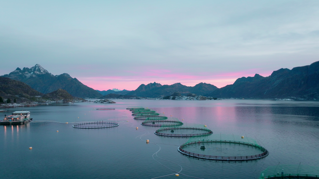
Ellingsen Seafood is a Norway-based producer of salmon. In the past, they have had issues with heart and skeletal muscle inflammation (HSMI) at their production plant in Ofotfjorden in Nordland. From May 2020 to May 2021, Ellingsen and Skretting Norway embarked on a health monitoring project to see if the use of a nutritional solution could support the fish during a HSMI outbreak and reduce the extent of it.
‘I wanted to test a feed with high omega-3 content, since we know that this is beneficial for the fish during an outbreak,’ says Steinar Myrvold, a veterinarian at Ellingsen Seafood.
Two groups of fish were monitored closely – one which was PRV-1 positive, and the other which was PRV-1 negative. PRV is the virus which causes HSMI. Samples were taken from the fish and tests were conducted on a monthly basis, from the time they were put in the pen to the time of harvest.
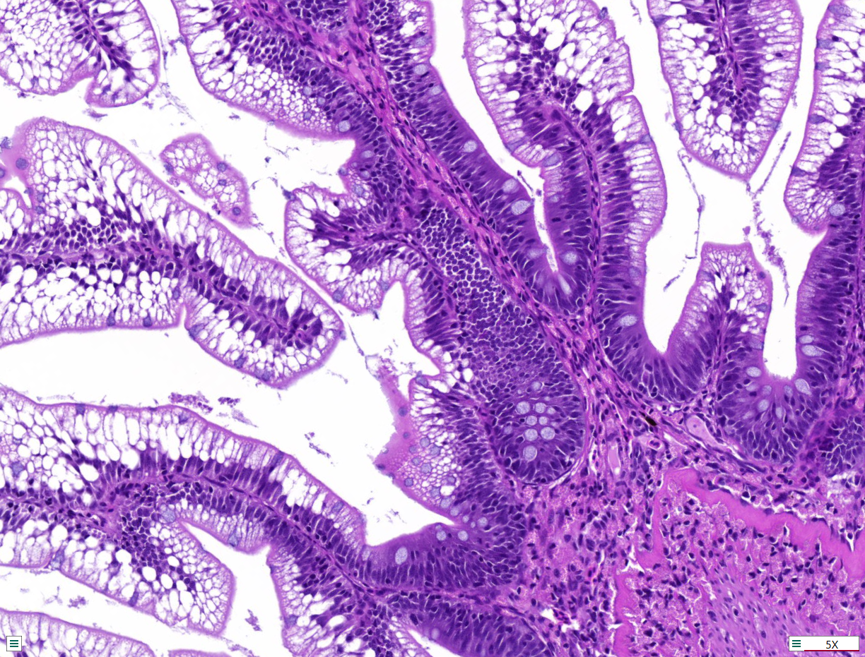
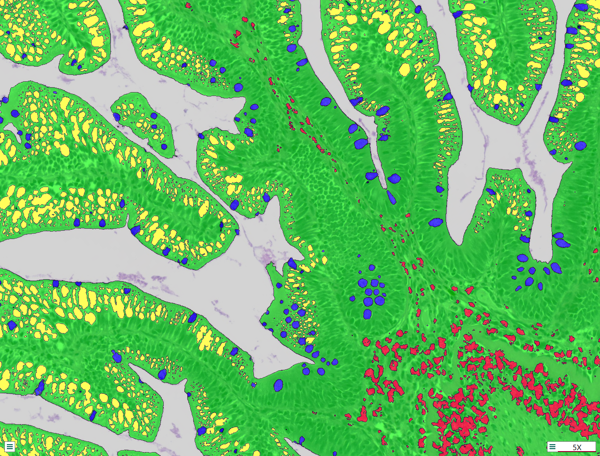
‘Together with Ellingsen and the Faculty of Veterinary Medicine at the Norwegian University of Life Sciences, we did several types of analysis for several months – blood chemistry, gene expression, pathogen detection, and histology. We looked at all the data combined to get an overview and understanding of what was going on with the fish,’ says Delphine Crappe, Team Lead Biotechnology and Manager at The Bubble. Production data was compared and evaluated as well.
The PRV-negative fish tested positive in August 2020, three months after the project kicked off. One month later, in September, results showed a further decline. However, there were no indications from the production data, nor from appetite and mortality, which would indicate that an HSMI outbreak was imminent. In such a scenario, production would normally have proceeded as usual, without any action taken.
‘Thanks to close collaboration with NMBU and Ellingsen Seafood and the fact that we acted quickly, we were able to take the appropriate measures and initiate a feeding program to support the fish during the HSMI outbreak,’ says Johan Rennemo, veterinarian in Skretting Norway. According to Myrvold, the result of this well-timed intervention was a milder outbreak of disease in the group that was PRV-1 positive.


Health monitoring projects like this are intended to complement, not replace existing conventional approaches to fish health. Their power lies in the fact that they can equip fish farmers with knowledge on how to prevent an outbreak of disease – specifically which feed to use, when to use it, and on which group of fish.
‘We now possess a better understanding of how to be more precise in utilising functional feed in relation to a HSMI outbreak. Given that the diet includes a high level of marine oils, which are scarce and increases costs, it should only be used when necessary,’ says Mads Martinsen, Director of Product Development and Sustainability in Skretting Norway. Both companies have continued to collaborate on other projects since.
‘We’ve demonstrated that it’s possible to be one step ahead of HSMI, and we will continue working on targeted nutritional solutions that can help support fish with other diseases. In times of sickness these could have a major positive impact on animal welfare. The potential here is still untapped,’ says Rennemo.
A paper on this health monitoring project has been published in Veterinary Research. To read the full paper, click here
Microarrays are used to detect the expression of thousands of genes at the same time. Here, you can see some examples of what we use microarrays for in The Bubble. Possible future applications include finding new biomarkers, documenting the effect of new diets, and better understanding how genes are regulated in various biological processes, including development, disease and treatment response.
Study 50,000 Atlantic salmon genes in one go

Study 27,000 whiteleg shrimp genes in one go

Study 25,000 sea bass and bream genes in one go

Study 11 tissues: skin, hepatopancreas, head kidney, heart, liver, fin, pleopods, gills, intestine, muscle, spleen

Examine the effect of protein- and fat-rich diets through observing pathway changes and gene expression levels


Our biotechnology specialists have been working with cell culture at The Bubble since February 2022, and are motivated by the principle of the 3Rs: Replace, Reduce, and Refine. By using cells, we are able to replace and reduce the use of live animals in the lab.
The team works with immortalised cells from fish to help develop anti-inflammatory products, amongst others. The cells are maintained in a growing medium at a constant temperature, on average doubling their numbers every week, making this an efficient and cost-effective method for research. Primary cells are also extracted from various fish organs in order to cultivate them. Within a few hours we are able to obtain millions of cells from each tissue, providing excellent models for health or growth studies within aquaculture.
Cell culture will play an increasingly important role in the early stages of product development. In a recent experiment, muscle cells were isolated, and their growth rate measured. In the future, this could be applied to the study of new grower products for aquaculture species.