Kýries asthéneies se ektrefómena thalássia eídi
![]()
![]()
-
Brain
-
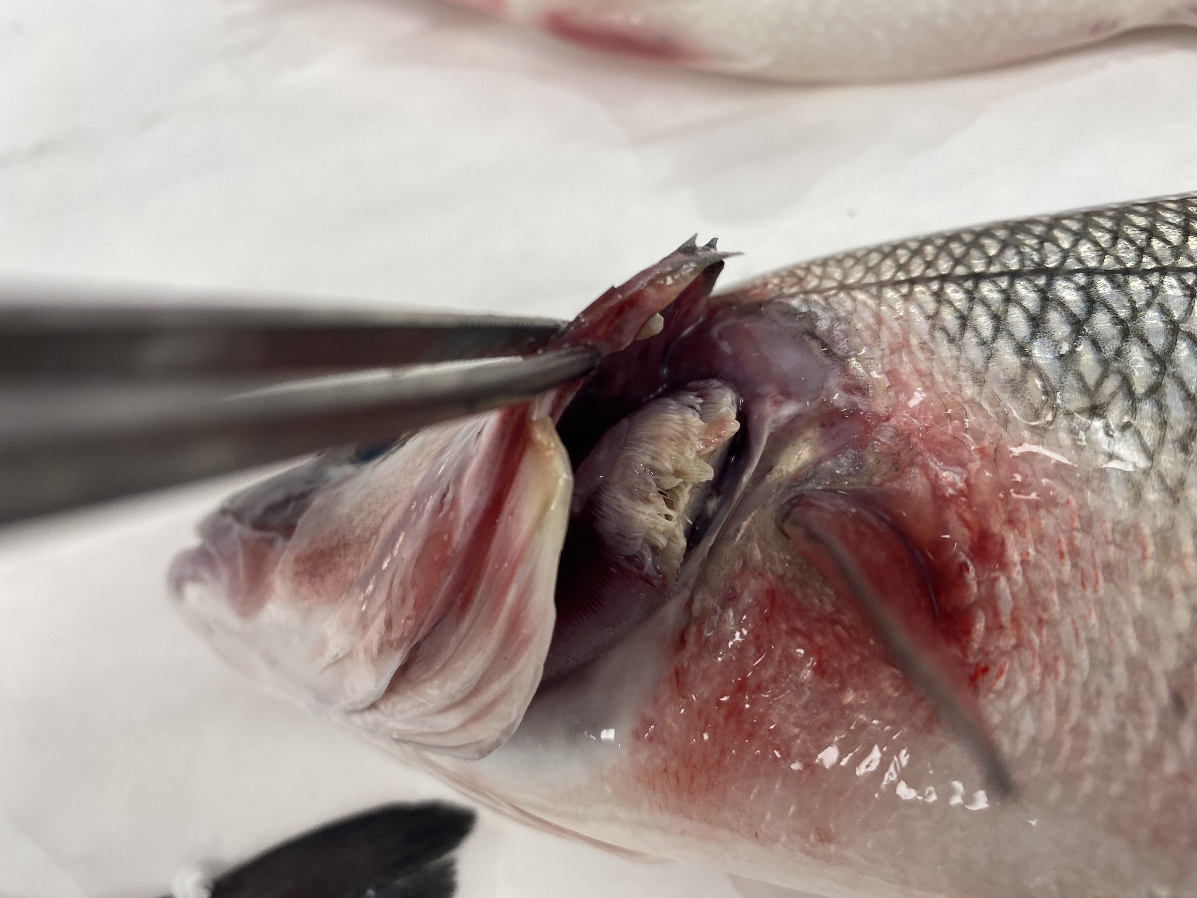
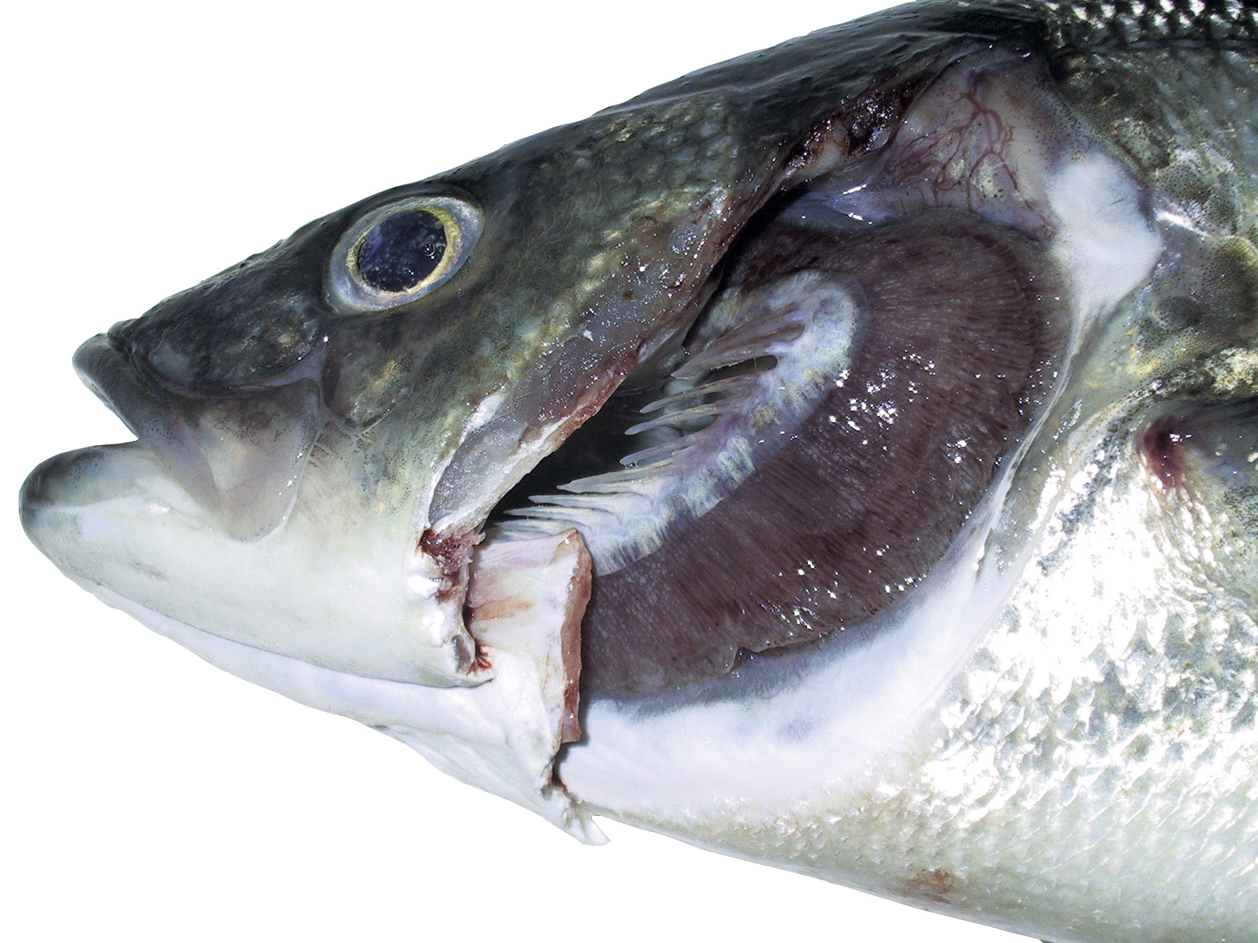
Gills
-
Heart
-
Liver
-
Spinal cord
-
Gall bladder
-
Swim bladder
-
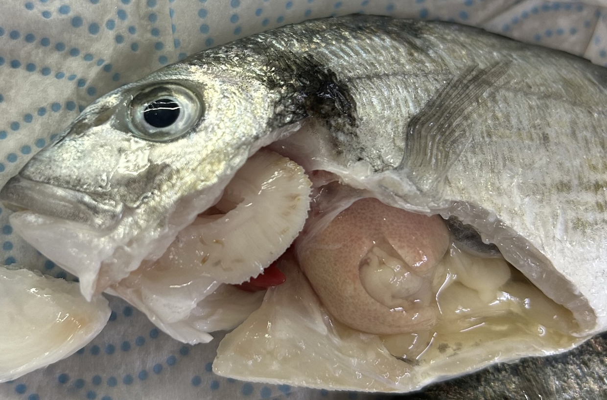
Spleen
-
Intestines
-
Stomach
-
Eggs
-
Vent
-
Kidney
-
Urinary bladder

Viral
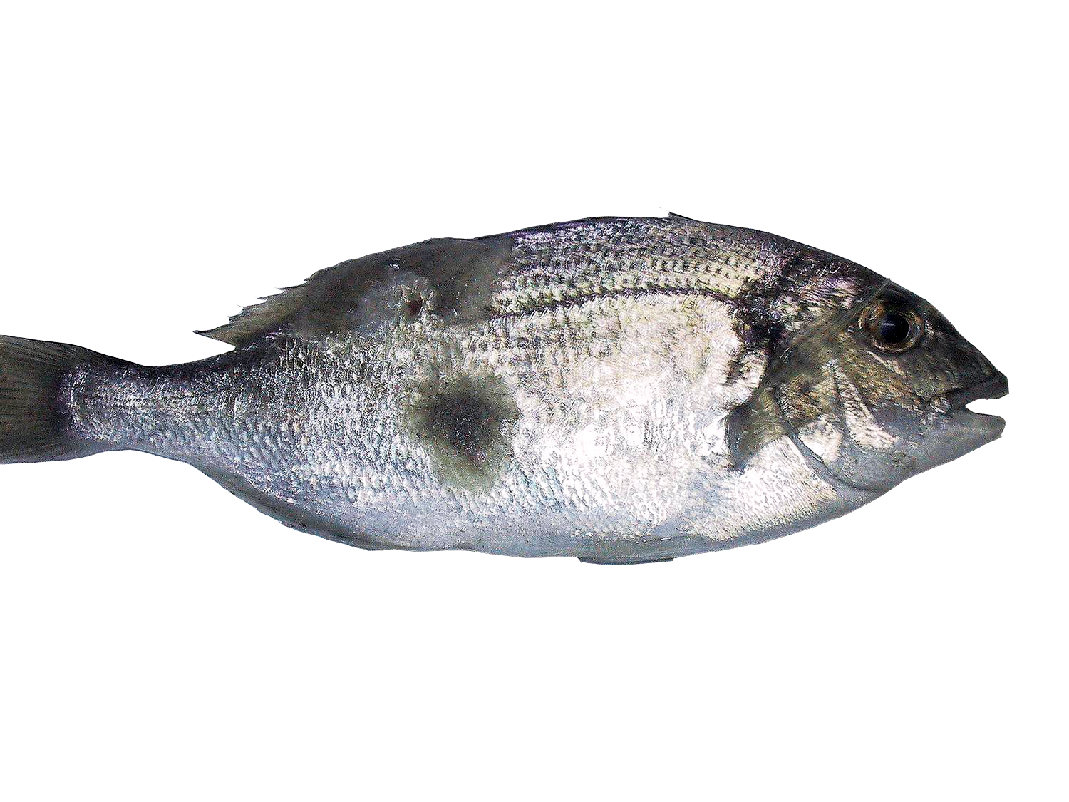
Lymphocystis
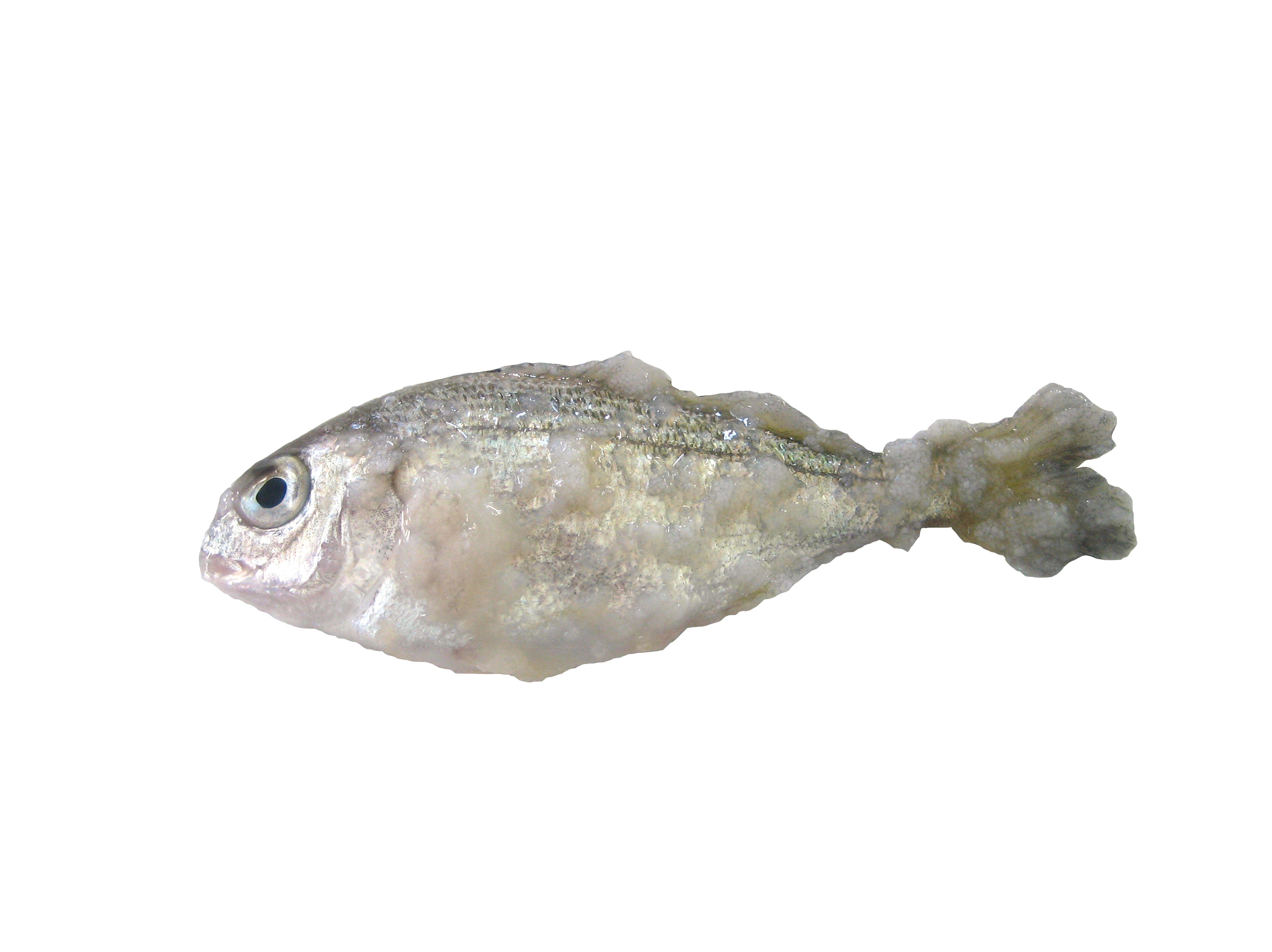
Agent: Lymphocystivirus (Iridoviridae).
Symptoms: Affects primary juvenile sea bream, causing white/gray nodules on the skin (hypertrophic fibroblasts due to viral replication). High morbidity but very
low mortality.
Control: No treatment; usually resolves on its own, minimize handling and stress
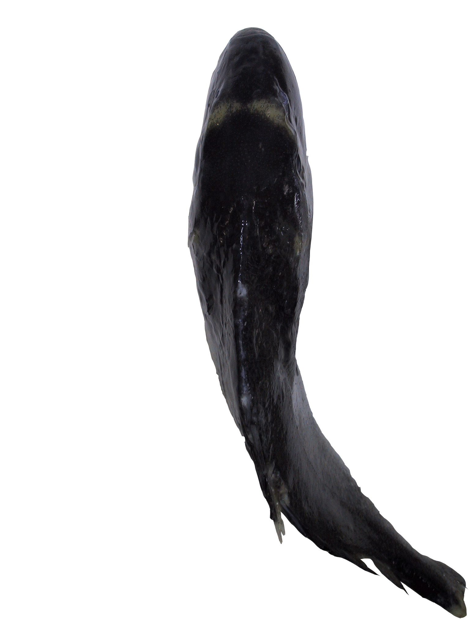
Viral Nervous Necrosis (VNN)
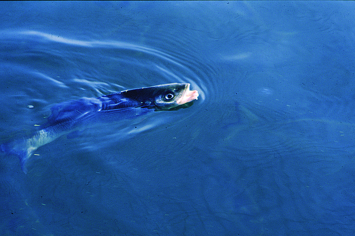
Agent: Nervous Necrosis Virus/ Nodavirus (Nodaviridae).
Symptoms: Affects sea bass (especially juveniles), sea bream and other species. It is a warm-water disease (≥18-20°C) with horizontal and vertical transmission. It causes erratic and altered swimming, brain congestion, swim bladder distension, brain congestion, and sometimes ulcerative lesions on the head. The mortality rate can reach the 100% in larvae. Adults may possibly be asymptomatic carriers.
Control: Prevention relies on vaccination, screening broodstock and using PCR diagnostics before entry of juveniles.
Fungal
Saprolegniasis
Agent: Saprolegnia spp. (Saprolegniales, Oomycete).
Symptoms: A common fungus in freshwater fish, it only affects gilthead sea bream in very low salinity conditions, such as during heavy rainfall in estuarine environments (pathological significance is minimal in marine species).
Control: Prevention involves minimizing the influx of freshwater during rains.
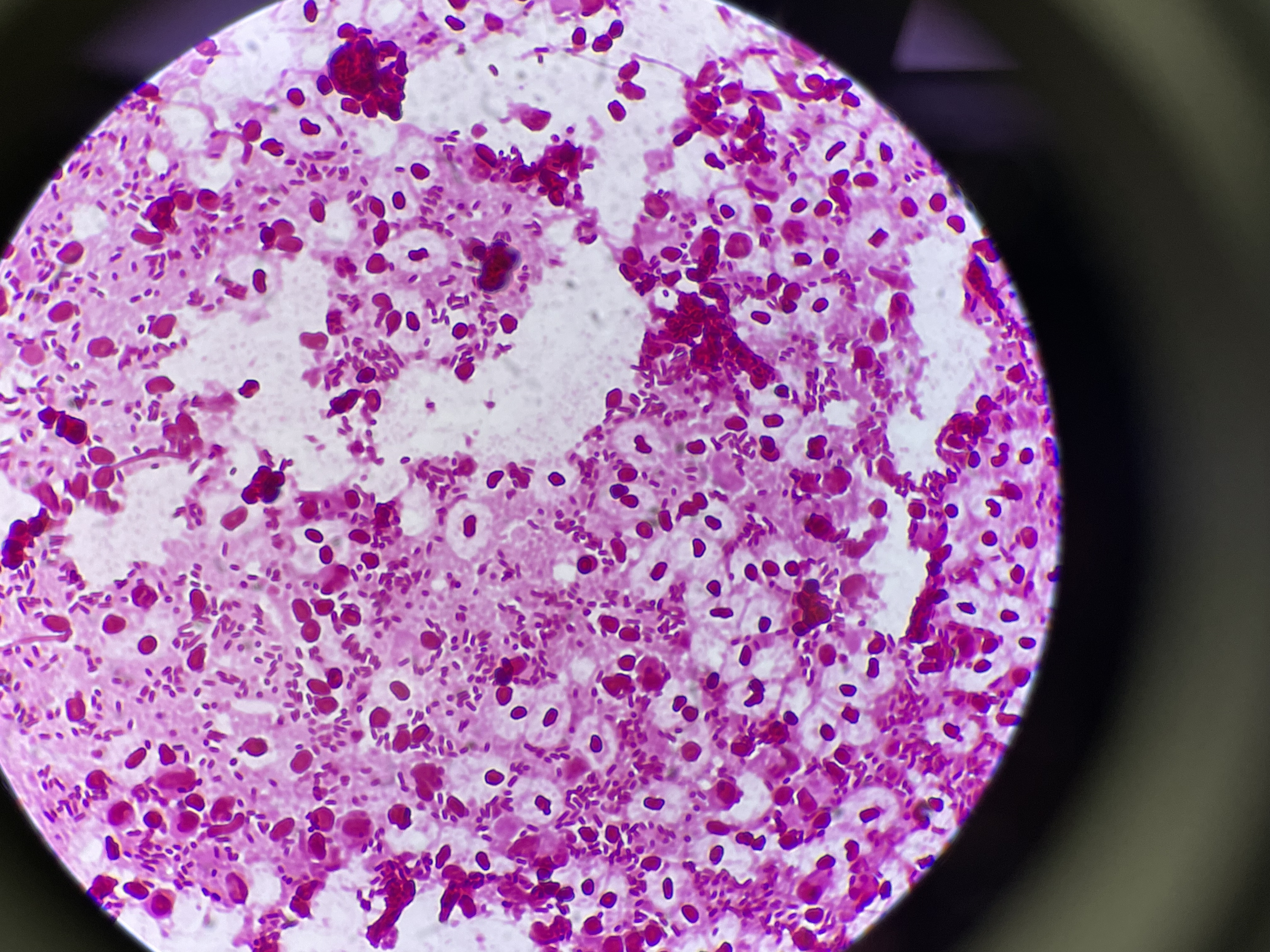
Bacterial
Lactococcosis
Agent: Lactococcus garvieae (Streptococcaceae, Gram-positive cocci).
Symptoms: It causes significant economic losses. Outbreaks curse with a septicemic-hemorrhagic infection with high morbidity and very high mortality in grow-out stages. Internal lesions include hemorrhages, exophthalmia, erratic swimming and darkening. Common in warm water (>15°C).
Control: Prevention relies on vaccination. Antibiotic therapy response is variable.
Vibriosis
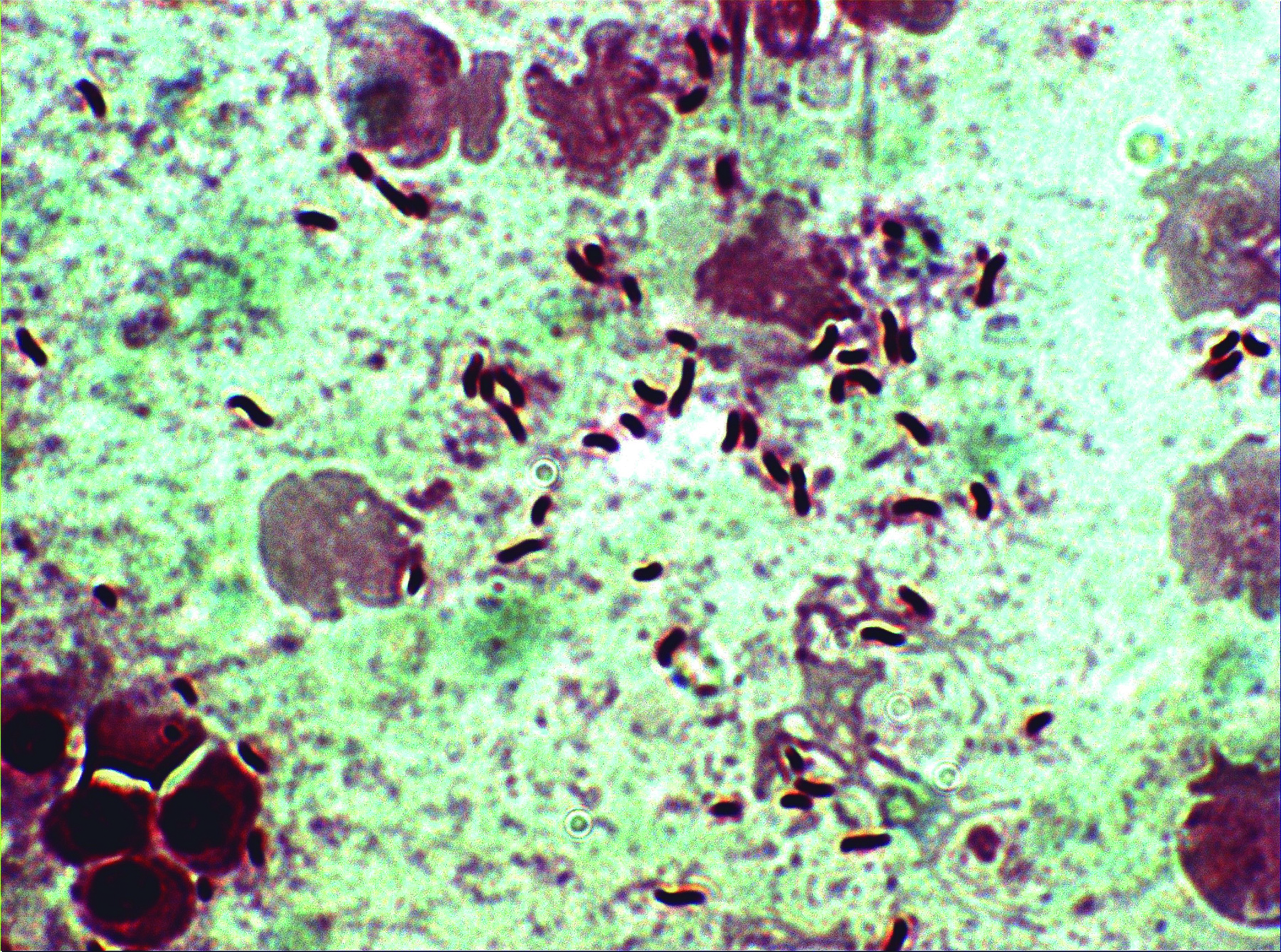
Agent: Vibrio anguillarum, Vibrio harveyi (Vibrionaceae, Gram-negative rod-shaped bacilli).
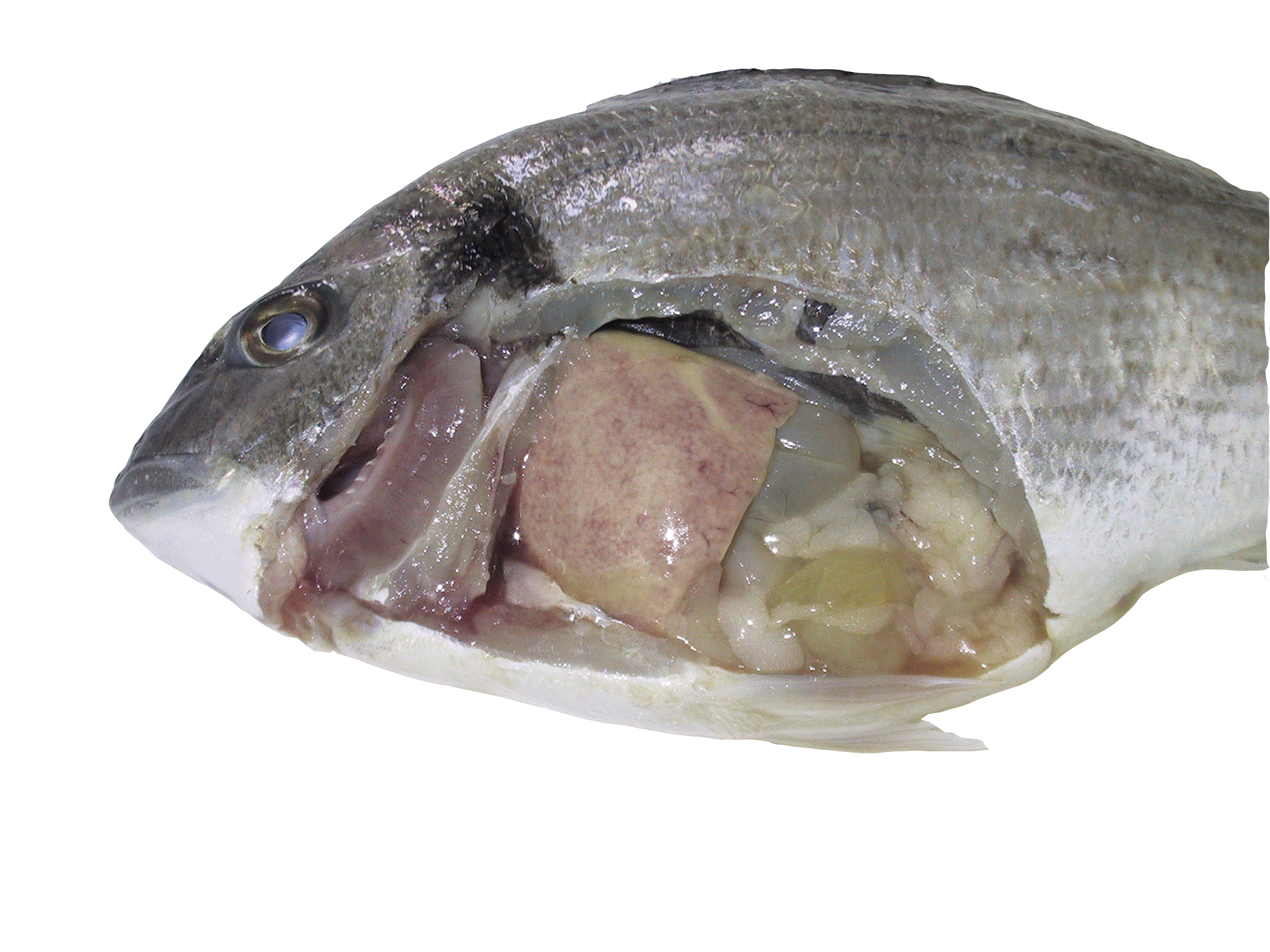
Symptoms: A warm-water disease exacerbated by temperature fluctuations. Spread through seawater. Lesions include splenomegaly, external and internal congestion and hemorrhages in the skin and perivisceral fat.
Control: Oral antibiotics and vaccination are effective.
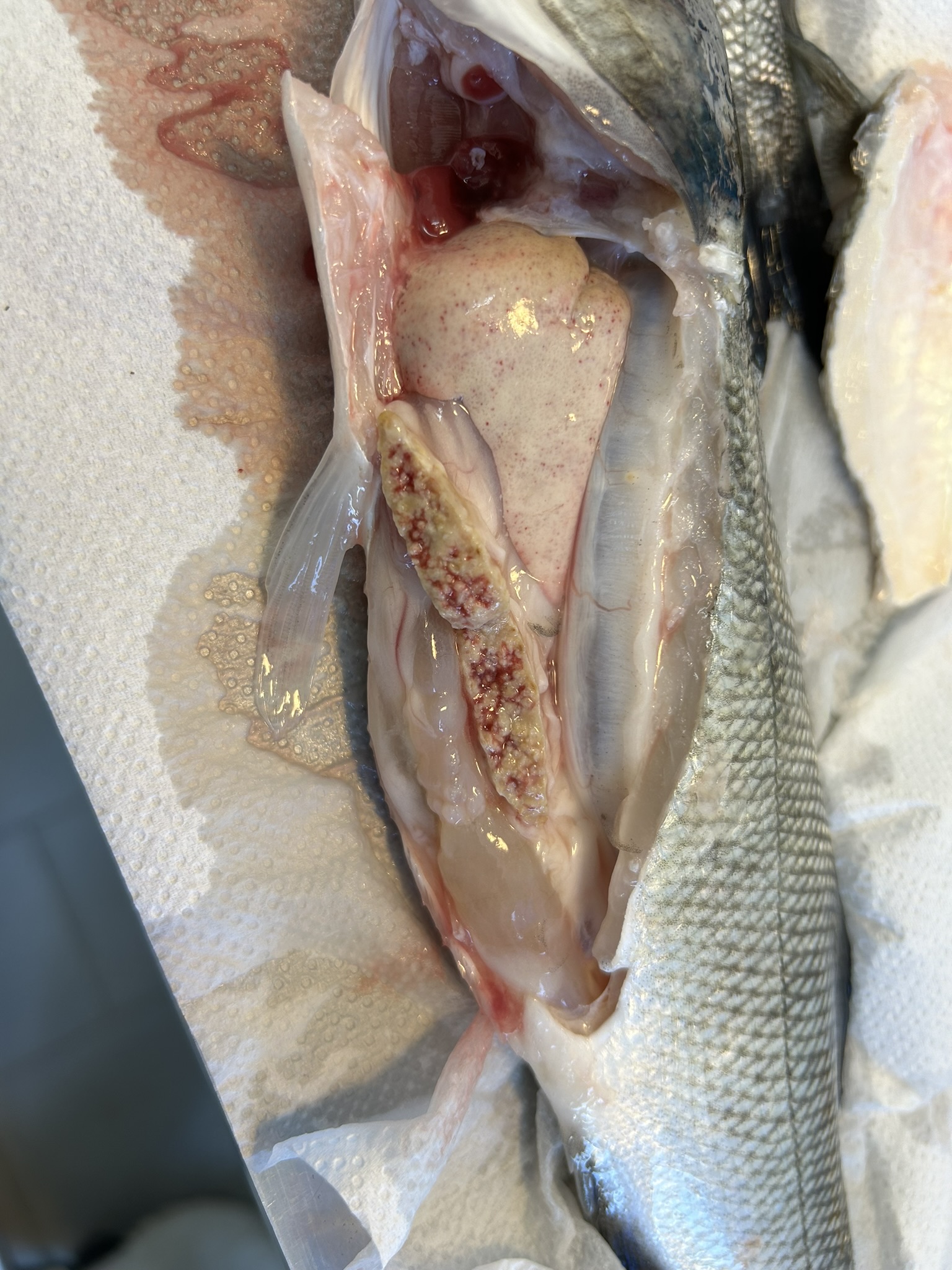
Nocardiosis
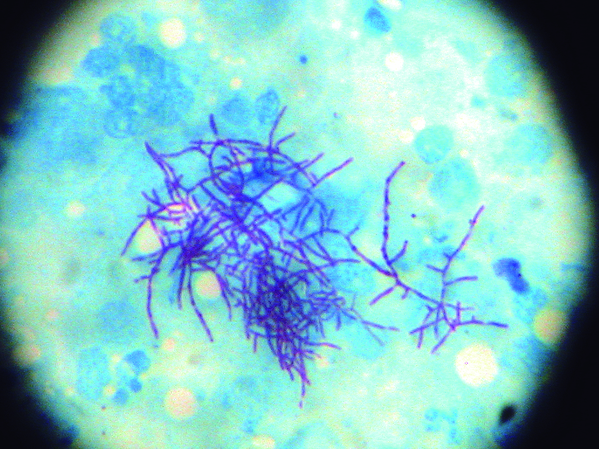
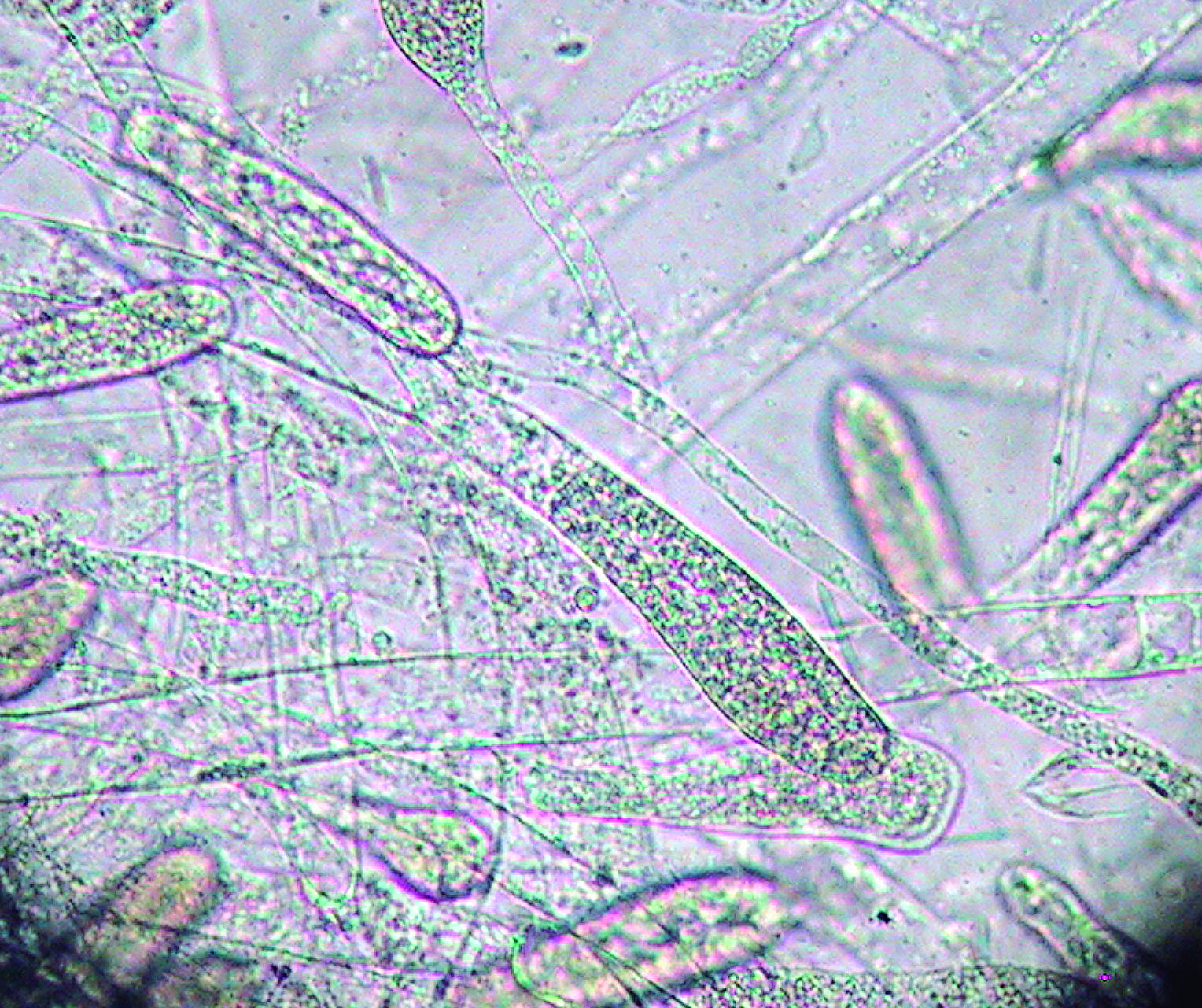
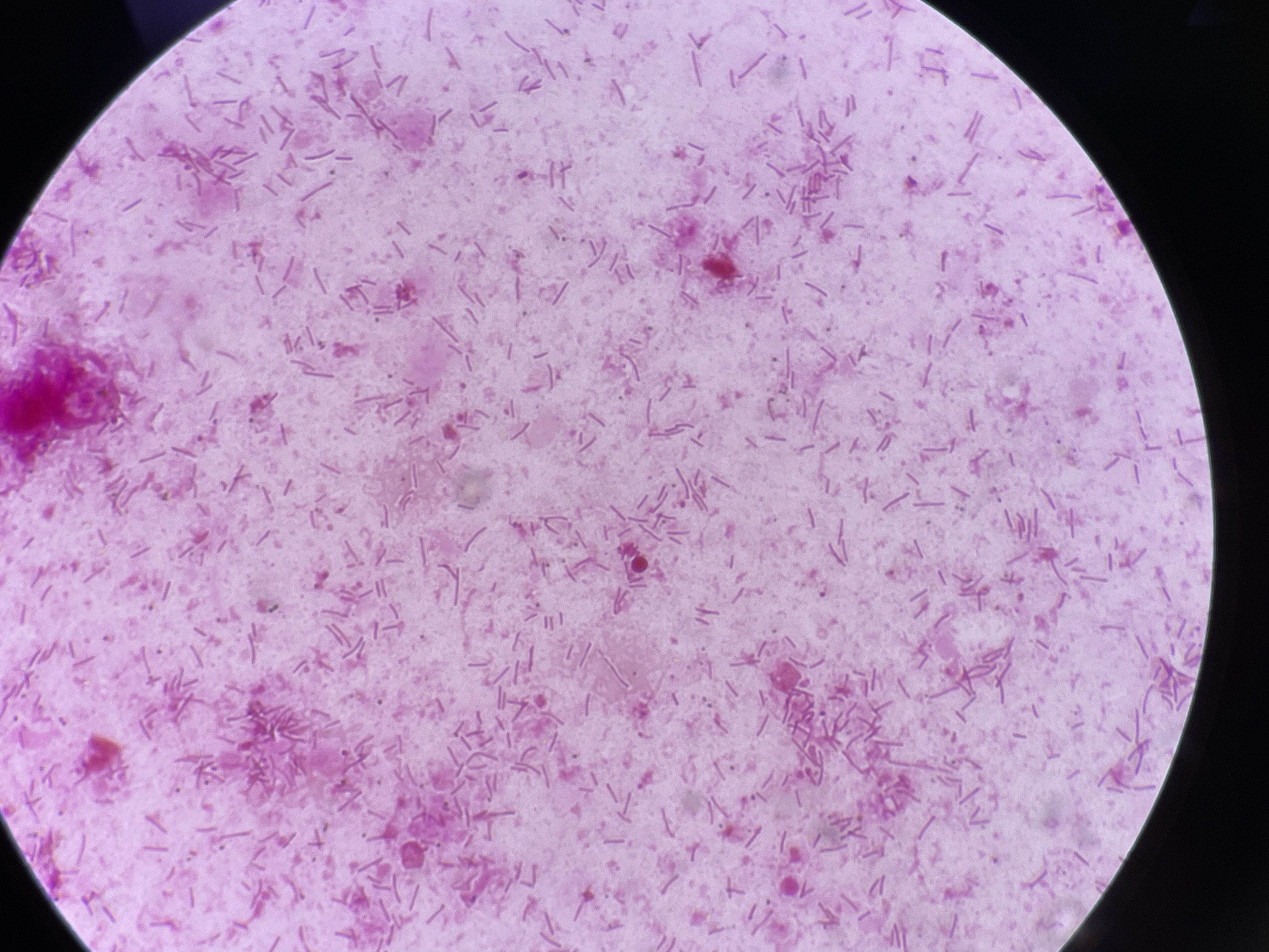
Agent: Nocardia seriolae and Nocardia spp. (Nocardiaceae, Gram-positive, partially Acid-alcohol resistant, branching and/or long filamentous bacteria).
Symptoms: A chronic disease characterized by ulcerative or caseous external lesions and granulomas in internal organs.
Control: Treatment with antibiotics is challenging, and affected fish should
be culled.
Mycobacteriosis
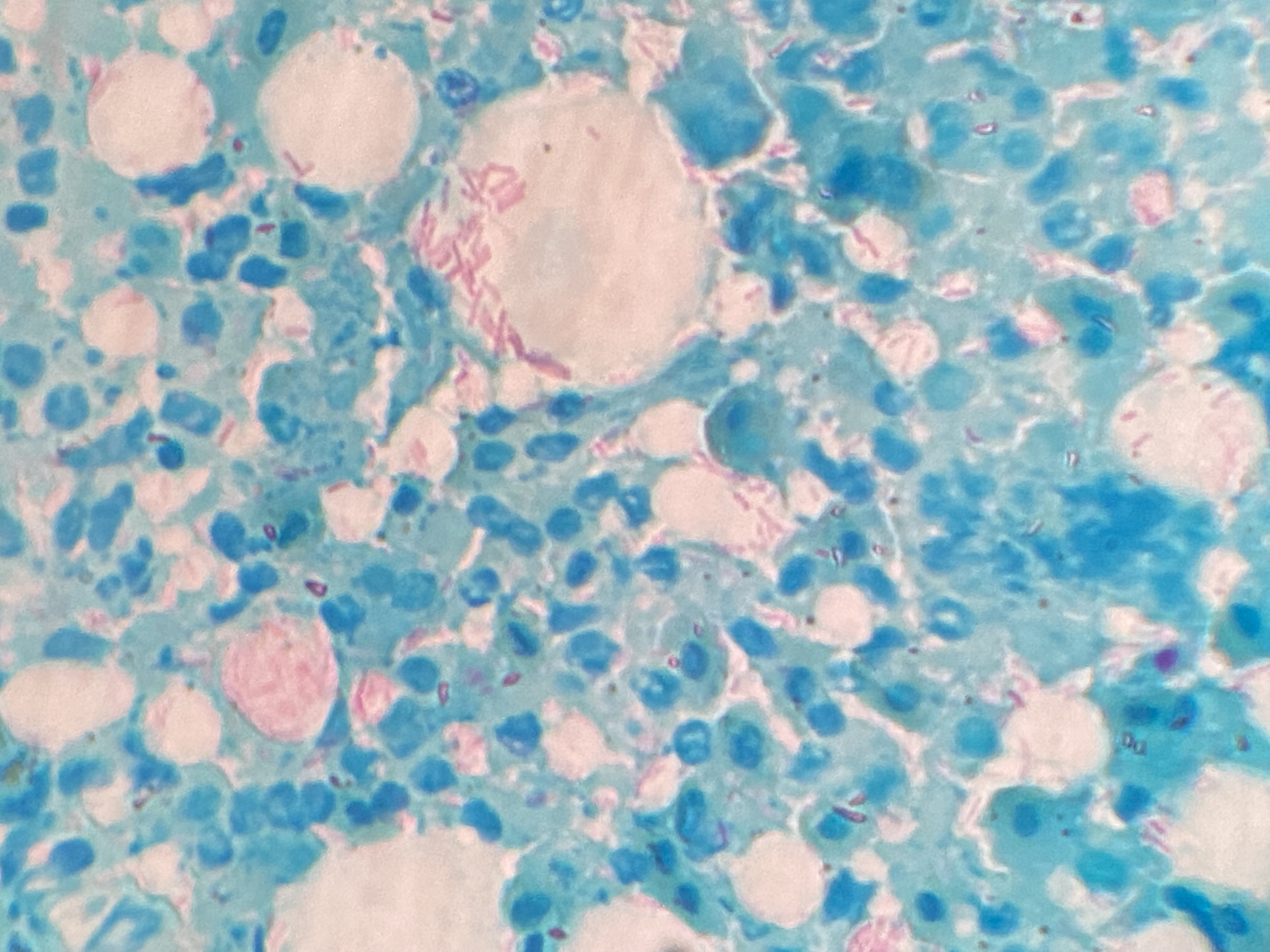
Agent: Mycobacterium spp. (Mycobacteriaceae, Acid-alcohol resistant Gram-positive rod-shaped small bacilli).
Symptoms: A chronic disease characterized by external hemorrhagic and ulcerative lesions, weight loss, and granulomas in internal organs. It occurs in warm water. Mycobacteria are zoonotic, so affected fish must be promptly removed. Some species of Mycobacterium are pigmented yellow or orange when cultured.
Control: Treatment is challenging, poor response to antibiotics.
Aeromoniasis
Agent: Aeromonas salmonicida subsp. salmonicida (Furunculosis), Aeromonas veronii and Aeromonas sobria (Aeromonadaceae, Gram-negative rod-shaped bacilli).
Symptoms: Symptoms include ventral congestion, hemorrhages, spleen inflammation, and perivisceral fat congestion.
Control: Antibiotic treatment is effective.
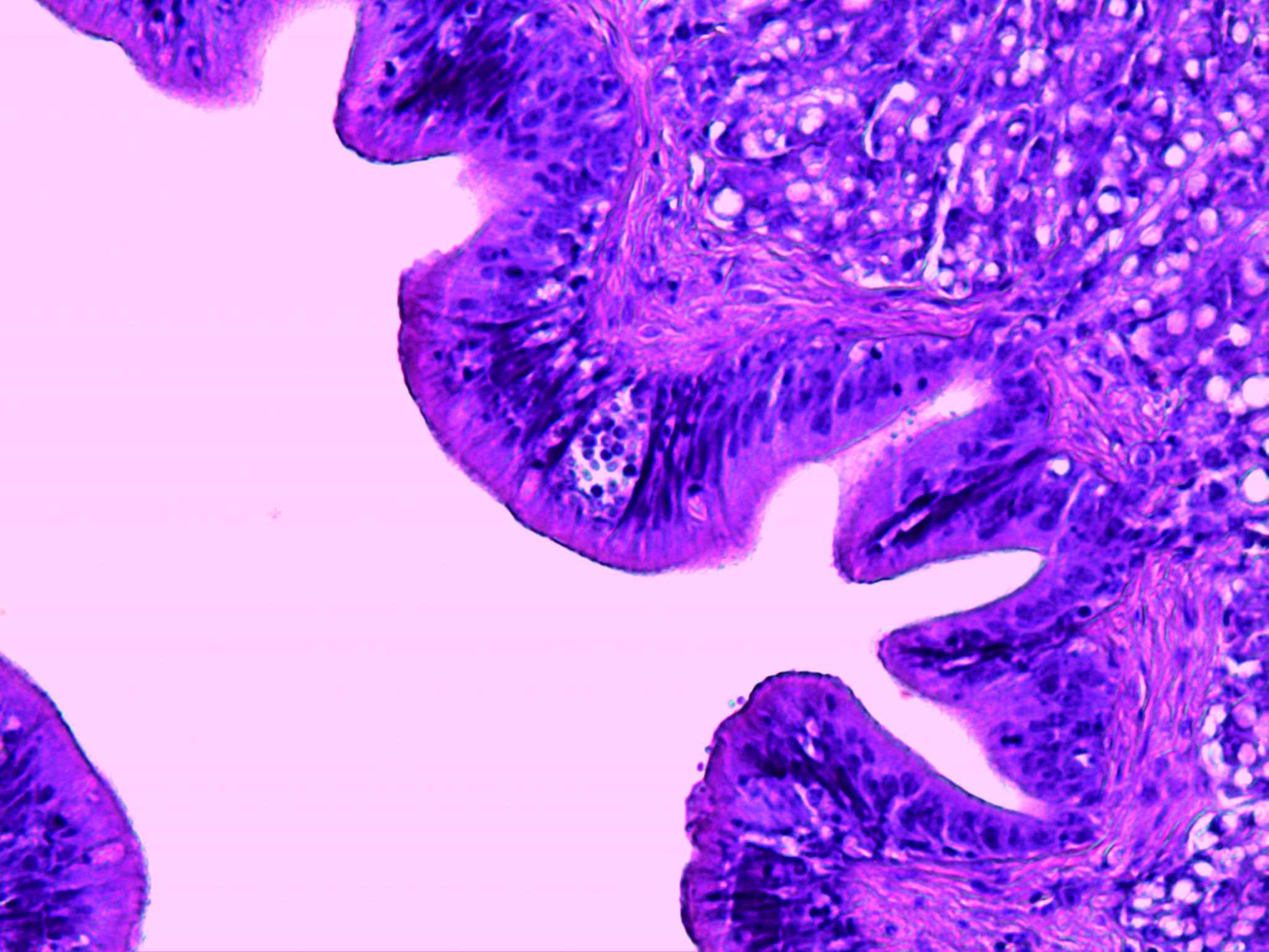
Epitheliocystis
Agent: Order Chlamydiales (Obligate intracellular bacterial pathogens). Traditionally, bacteria from the phylum Chlamydiae were the only known pathogenic agents, but etiology is now recognized as being more complex, including a range of Proteobacteria.
Symptoms: Sea bream and, less commonly, sea bass are affected. Caused by an intracellular bacterium that replicates within gill epithelial cells, leading to hypertrophy and that compromise gill function. Severe infections may cause hyperventilation and mortality.
Control: Antibiotic therapy response is variable. No vaccine is available.
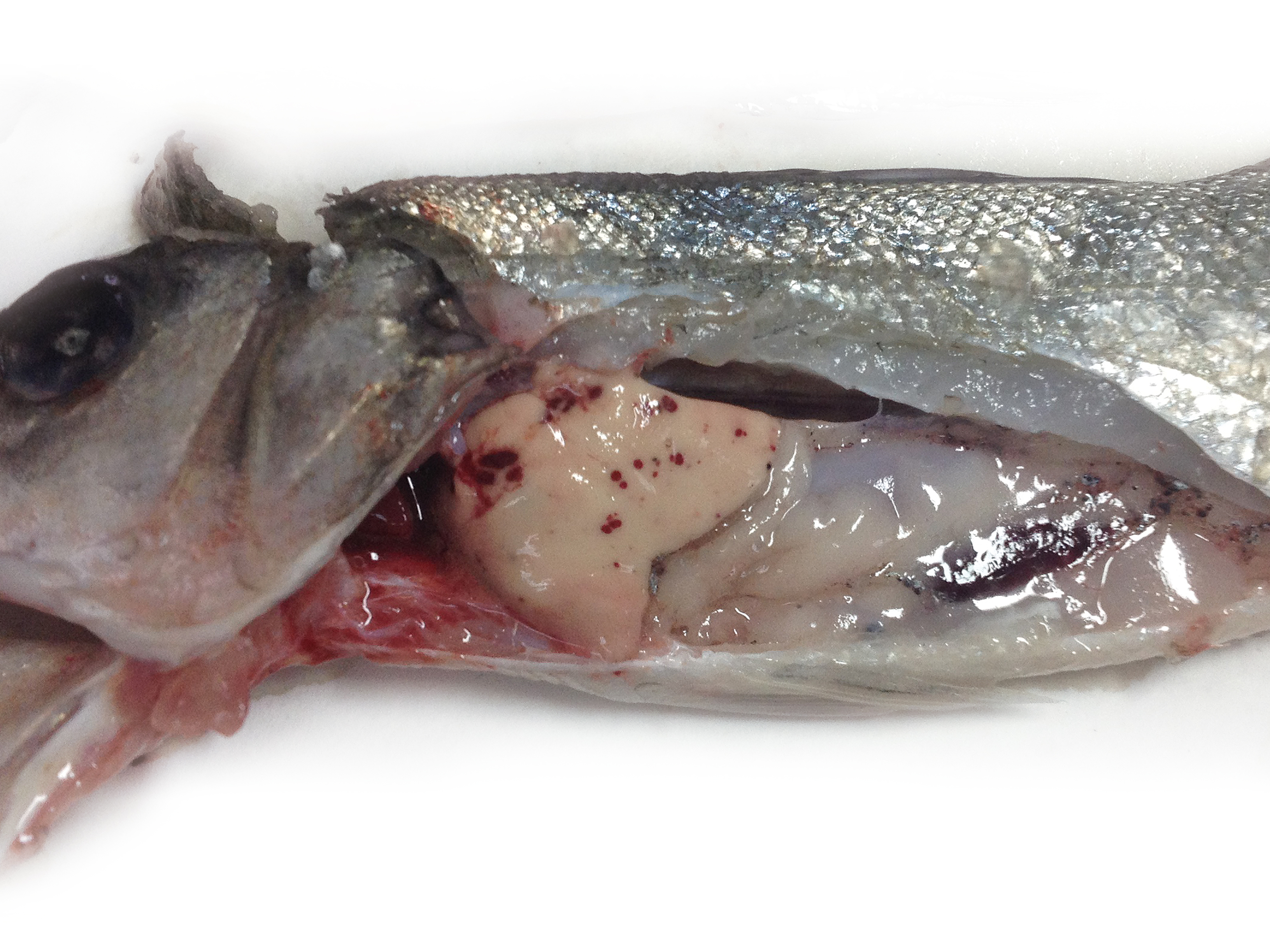
Photobacteriosis
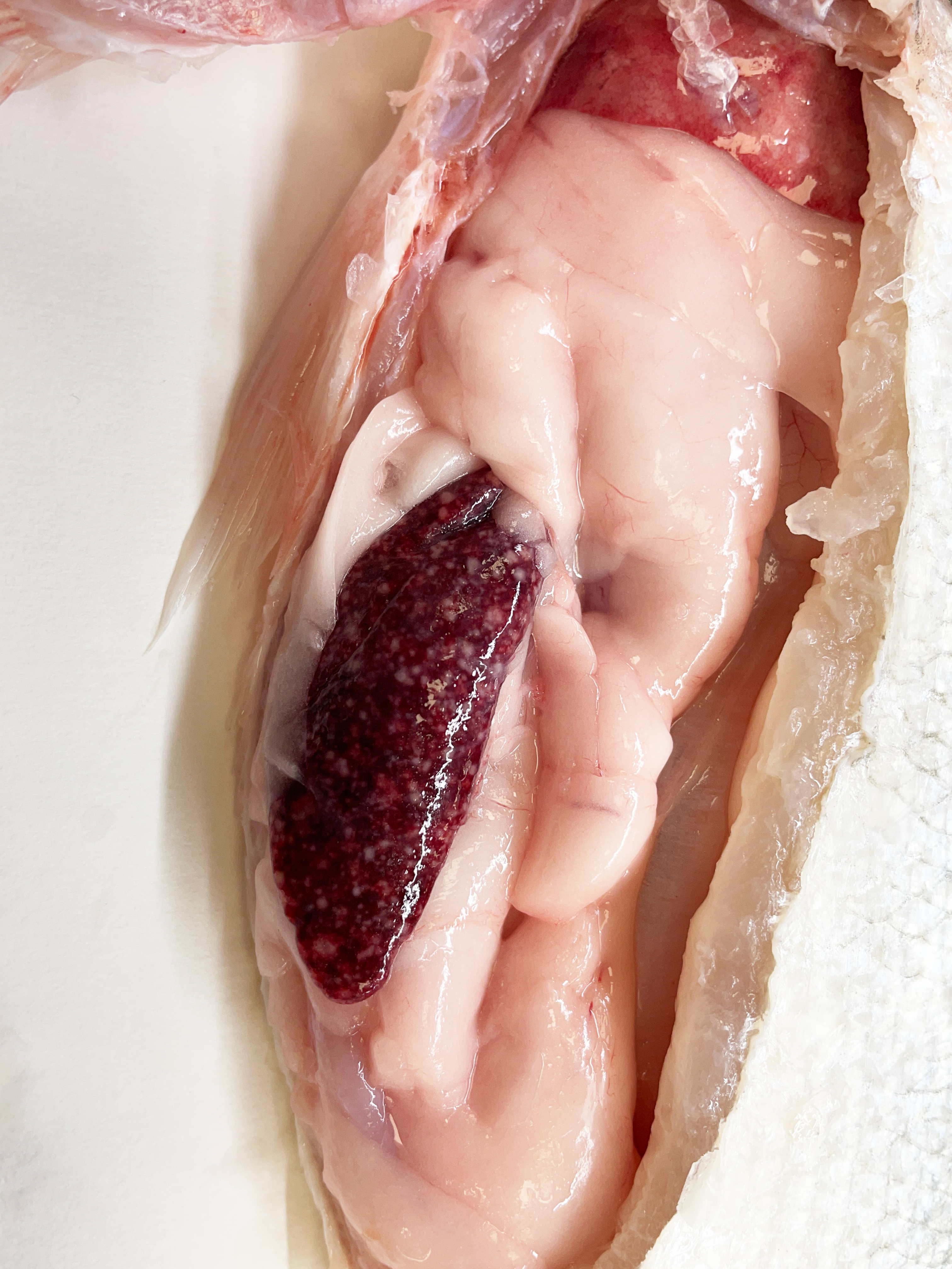
Agent: Photobacterium damselae subsp. piscicida (Vibrionaceae, Gram-negative rod-shaped bacilli).
Symptoms: Affects sea bass and sea bream. Common in warm water (>18-19ºC), outbreaks are normally linked to sudden temperature changes.
Lesions include intense spleen inflammation with granulomas. In hyperacute cases, splenic inflammation may be absent.
Control: Antibiotic treatment and vaccines are effective, although protection might not be enough in big fish (>2 years in water).
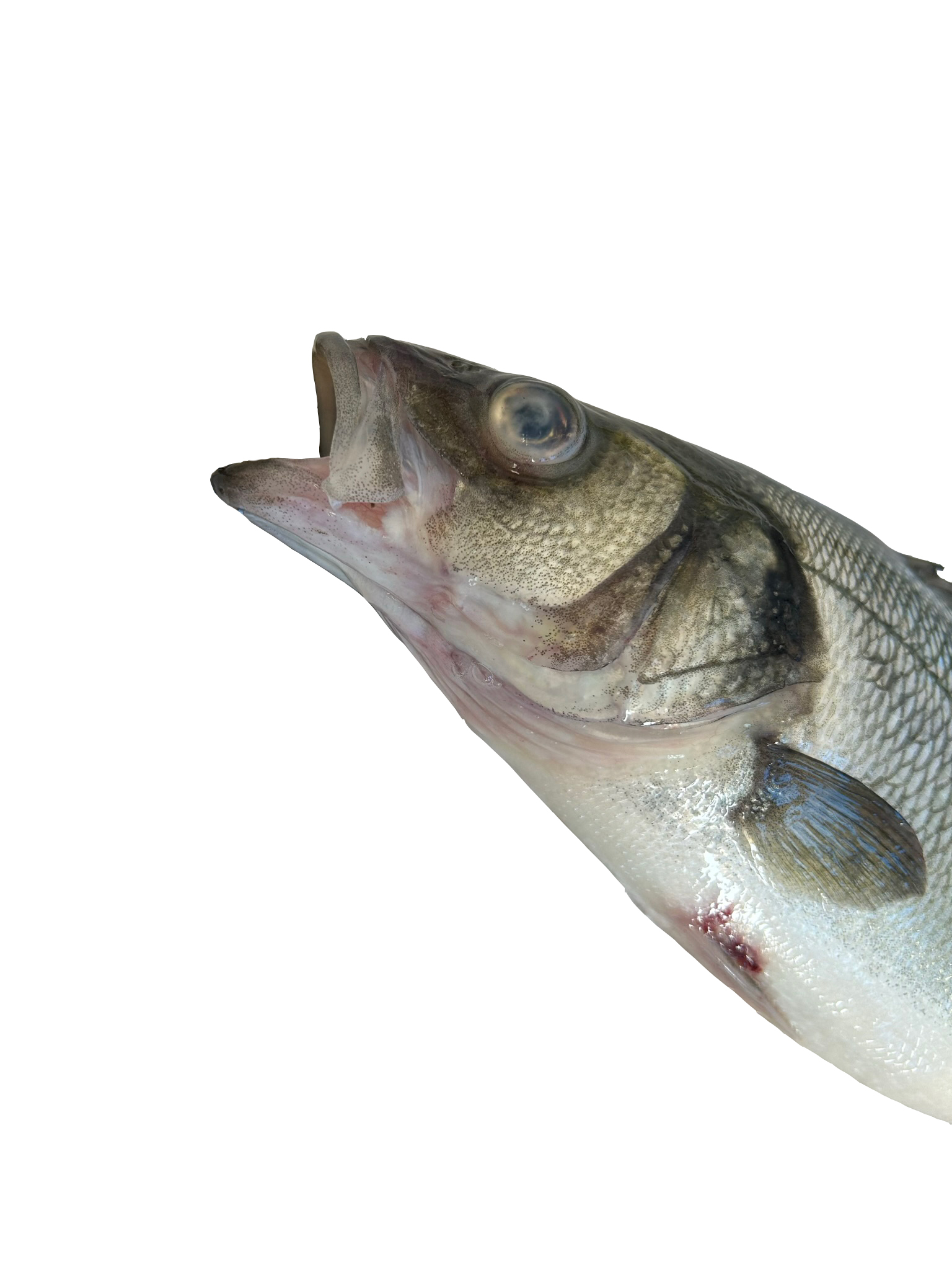
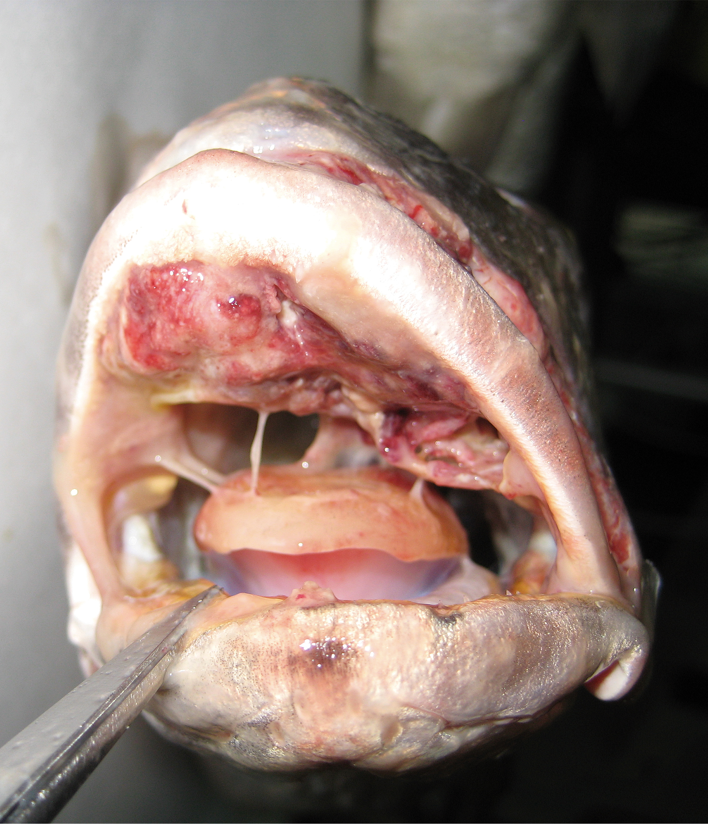
Tenacibaculosis
Agent: Tenacibaculum maritimum and Tenacibaculum spp. (Flavobacteriaceae, Gram-negative long slender rod-shaped bacilli).
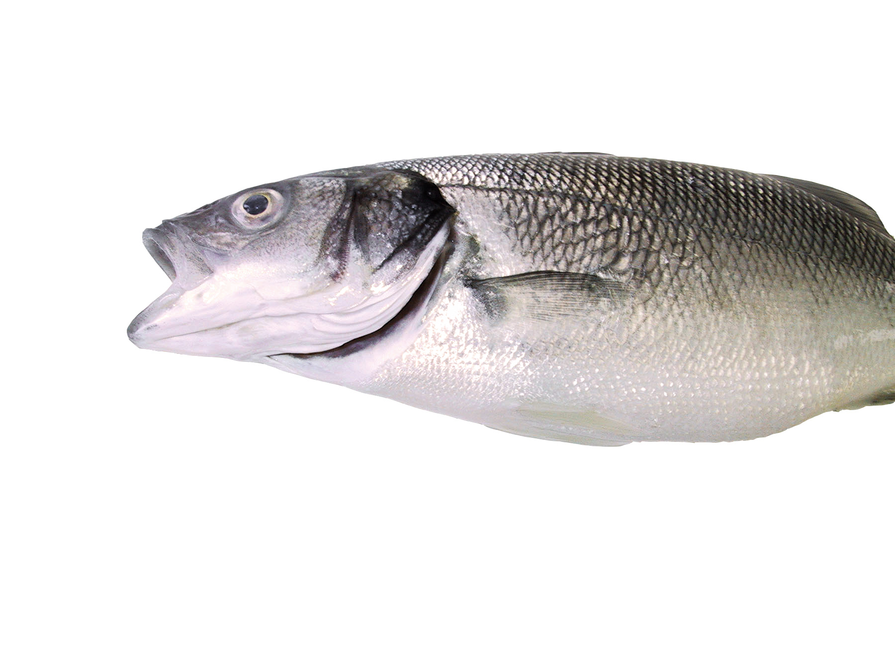
Symptoms: More frequent in winter and spring, this disease can occur anytime, as secondary contamination of skin erosions. Juvenile sea bream and sea bass exhibit several external clinical signs including mouth erosion, epidermal erosions and ulcers, fin necrosis, and tail rot. Larger fish may show necrotic yellowish gill tissue.
Control: Prevention includes reducing stress and vaccinating juveniles. Depending on the country regulation, it is possible to use external bath.
Parasitic
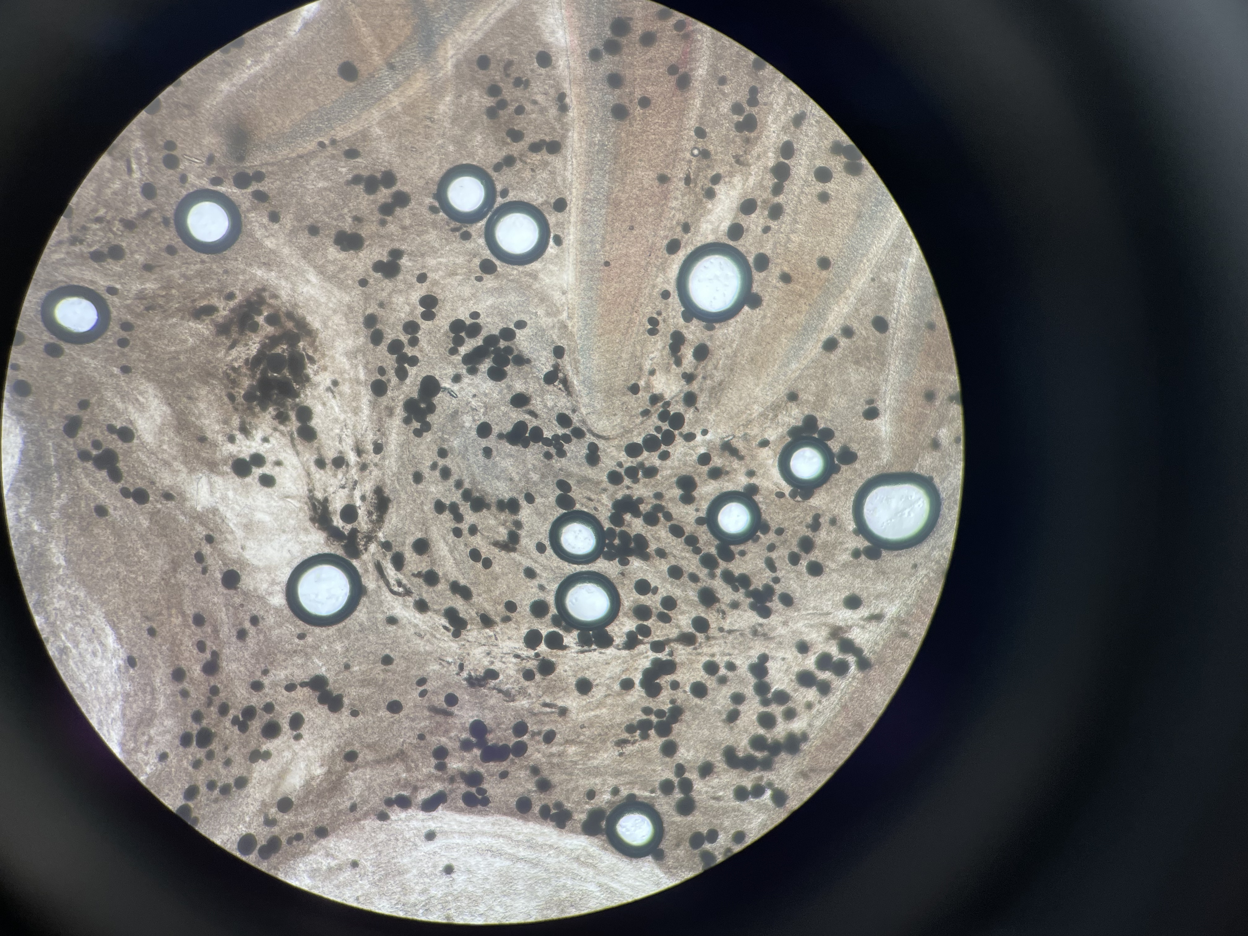
Cryptocarionosis
Agent: Cryptocaryon irritans (Cryptocaryonidae, Ciliate parasite).
Symptoms: Ciliated protozoan that forms visible whitish nodules on the skin and gills, primarily in summer. Horizontal transmission occurs fish-to-fish. The life cycle is influenced by temperature, salinity and photoperiod. Diagnosis involves observing trophonts or tomonts in skin and gill samples.
Control: Depending on the country regulation, it is possible to use external bath.
Monogenean Parasitosis
Agent: Furnestinia echeneis / Diplectanum aequans / Calceostoma sp. / Sparicotyle chrisophrii/ Sciaenocotyle pancerii.
Symptoms: Flatworm parasites found on fish gills (Gill flukes), feeding on mucus and blood. High infestations can cause respiratory issues and anemia, with increased mortality in spring and early summer.
Control: Depending on the country regulation, it is possible to use external bath or antiparasitic feed.
Copepod Crustacean Parasitosis
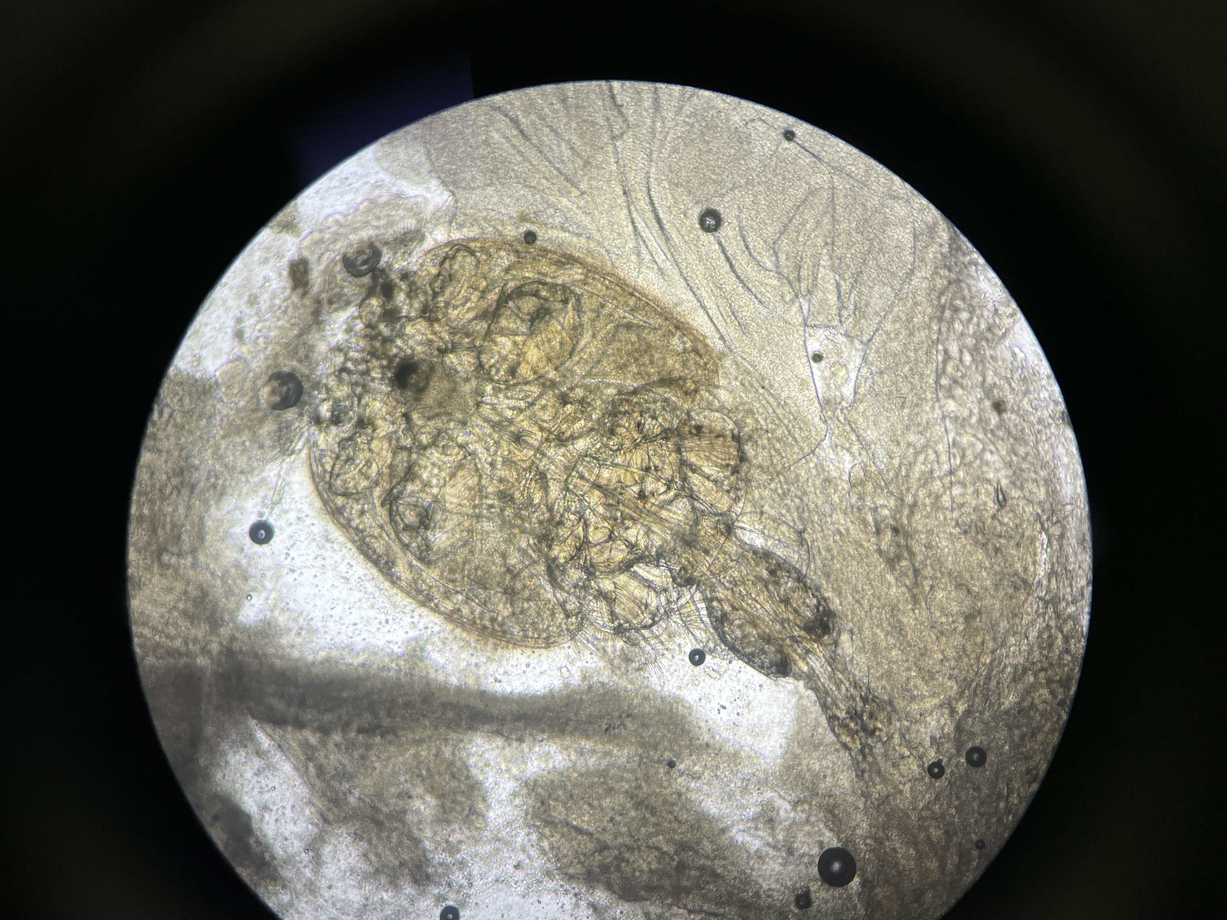
Agent: Caligus spp. and Lernanthropus kroyeri (Copepod crustacean parasite).
Symptoms: Metazoans that infest the oral cavity (Caligus spp.) and gills (L. kroyeri). Mortality is low, but they can reduce feeding efficiency.
Control: Depending on the country regulation, it is possible to use external bath or antiparasitic feed.
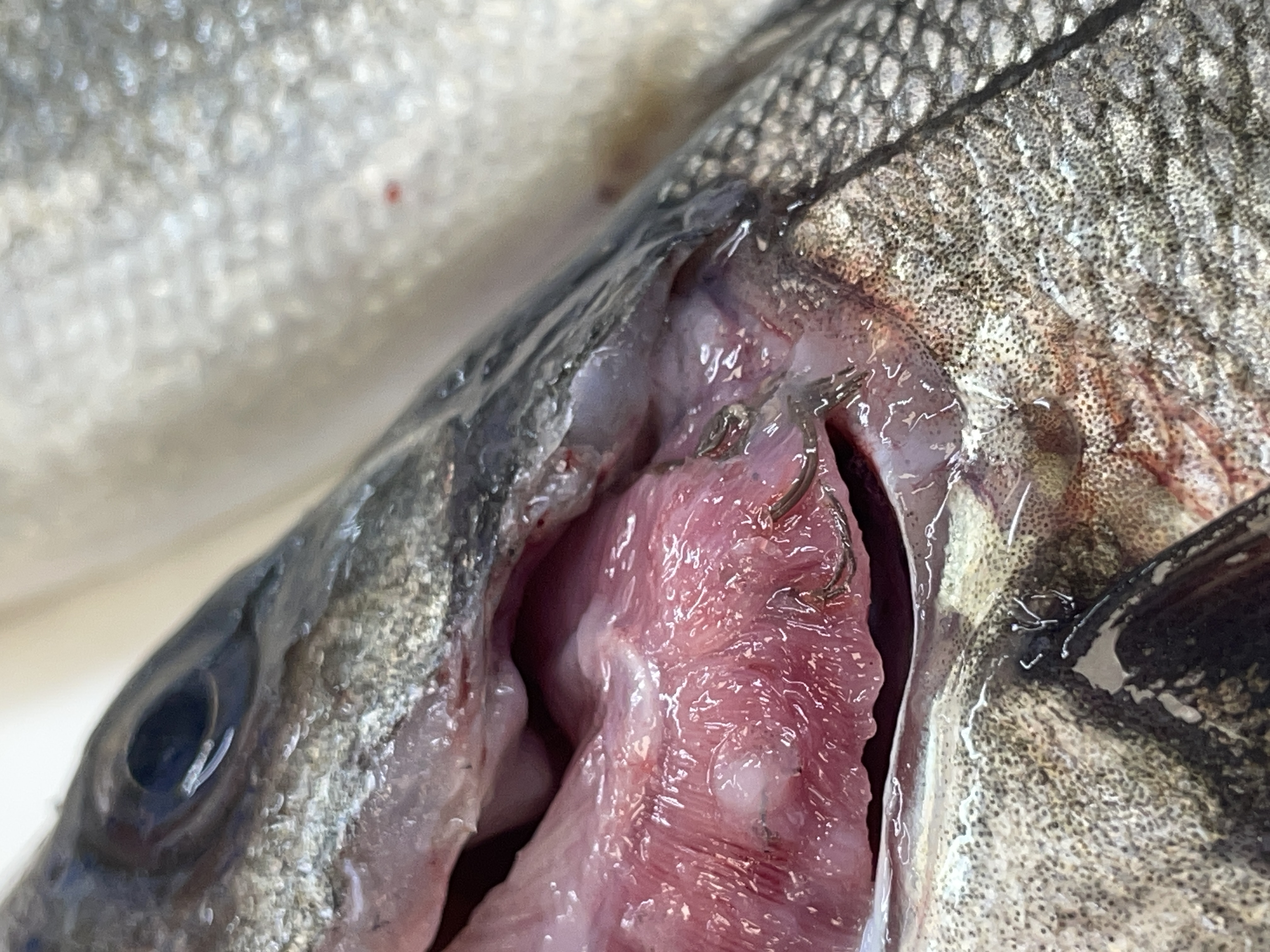
Isopod Crustacean Parasites
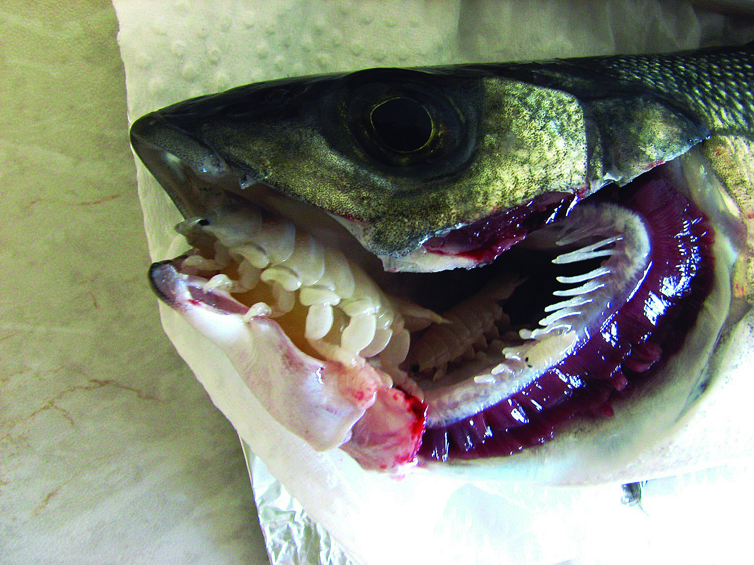
Agent: Ceratothoa spp., Nerocila spp., Anilocra spp. (Isopod crustacean parasite).
Symptoms: Parasitise the oral cavity (Ceratothoa spp.) or skin (Nerocila spp., Anilocra spp.) mainly in summer.
Control: Prevention involves separating fish of different sizes to reduce transmission.
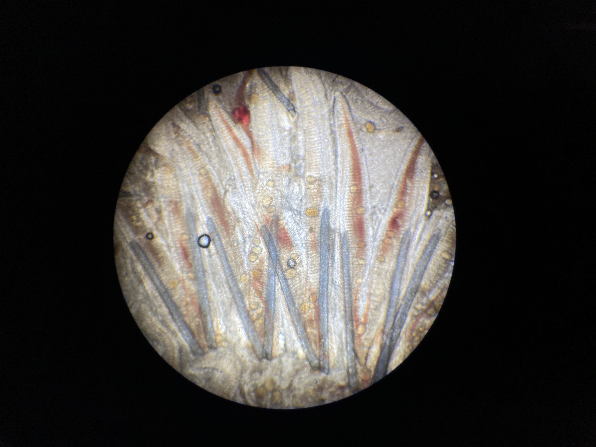
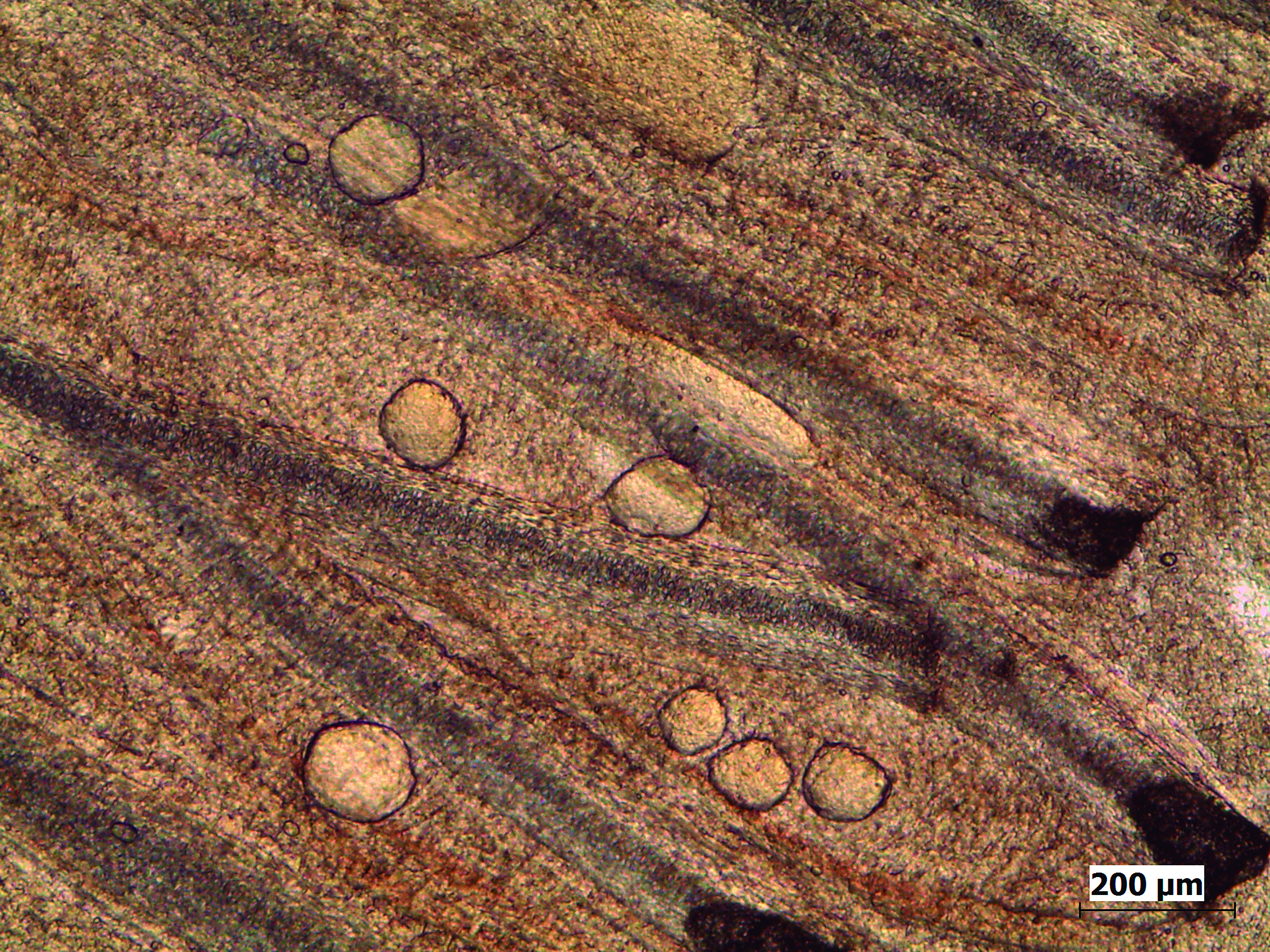
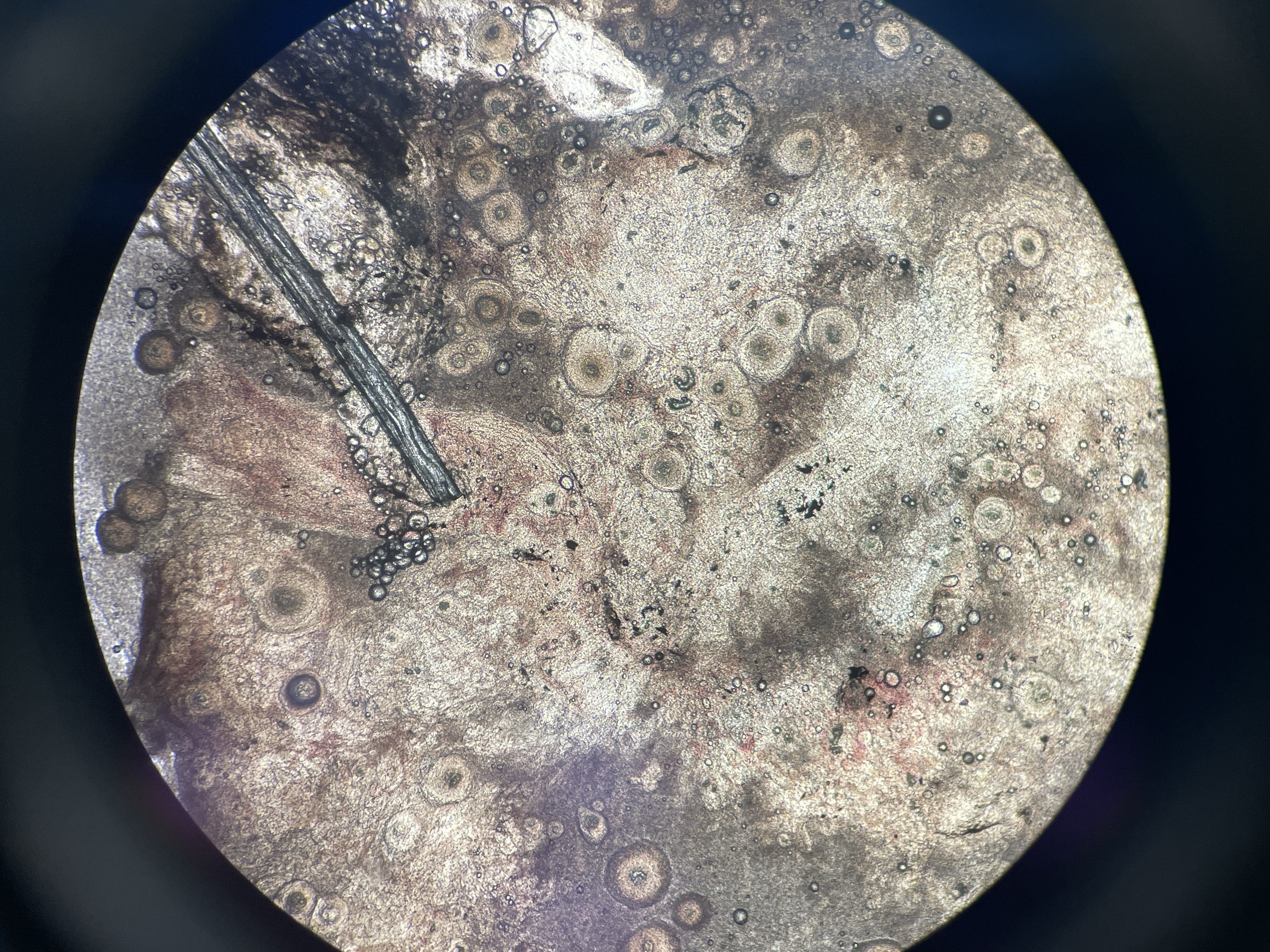
Trematode Digenean Parasitosis
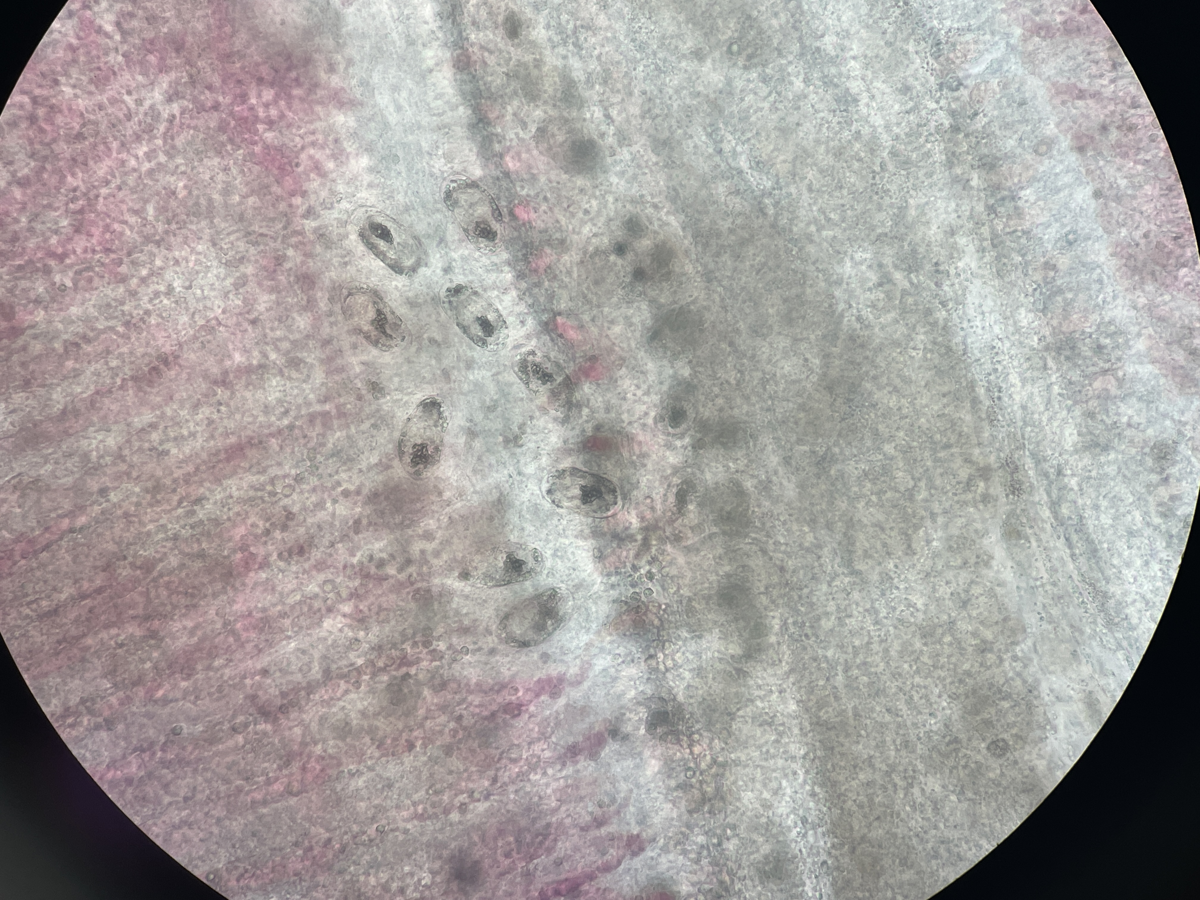
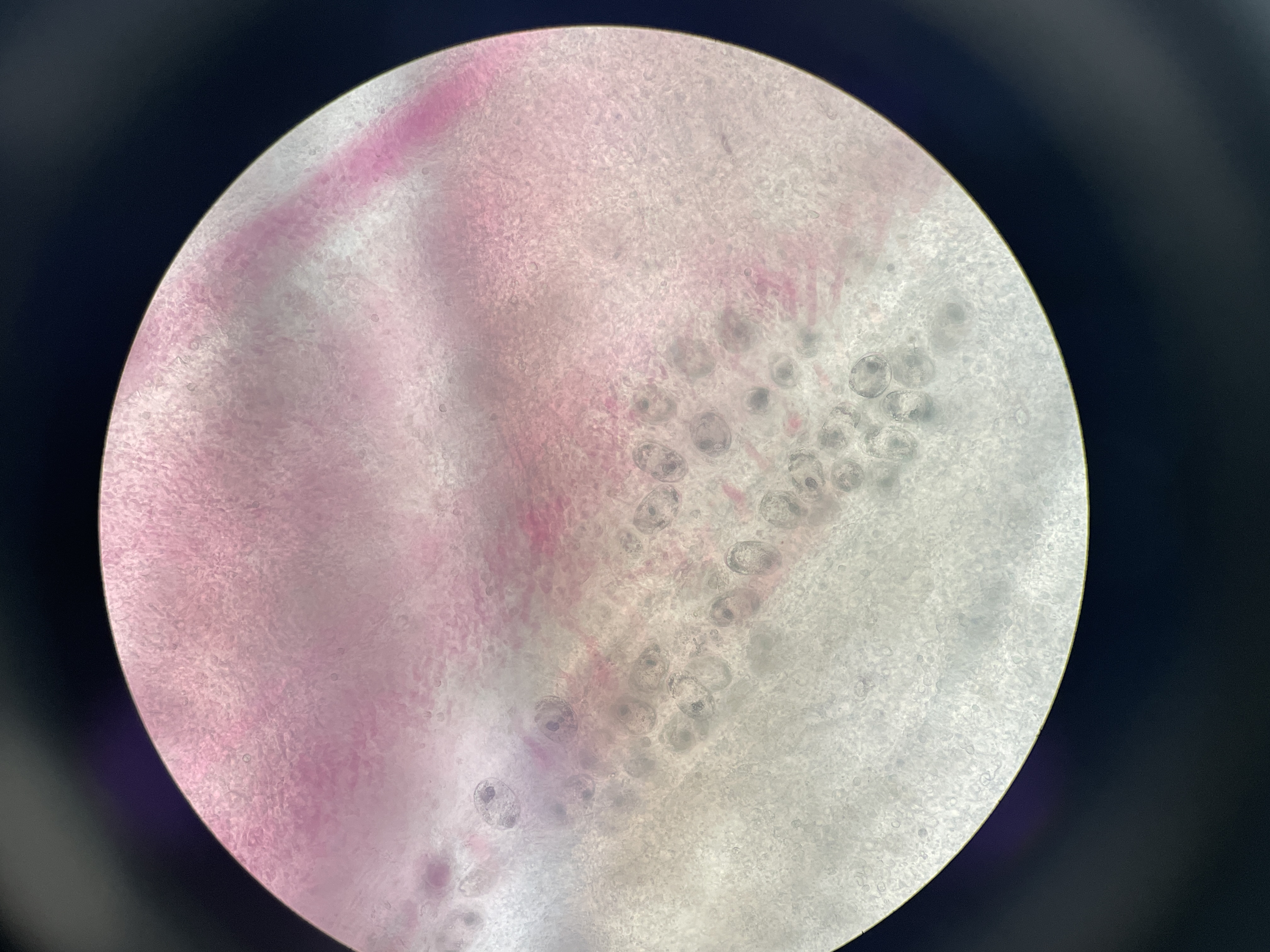
Agent: Cardicola aurata (Sanguinicolidae, Digenean parasite).
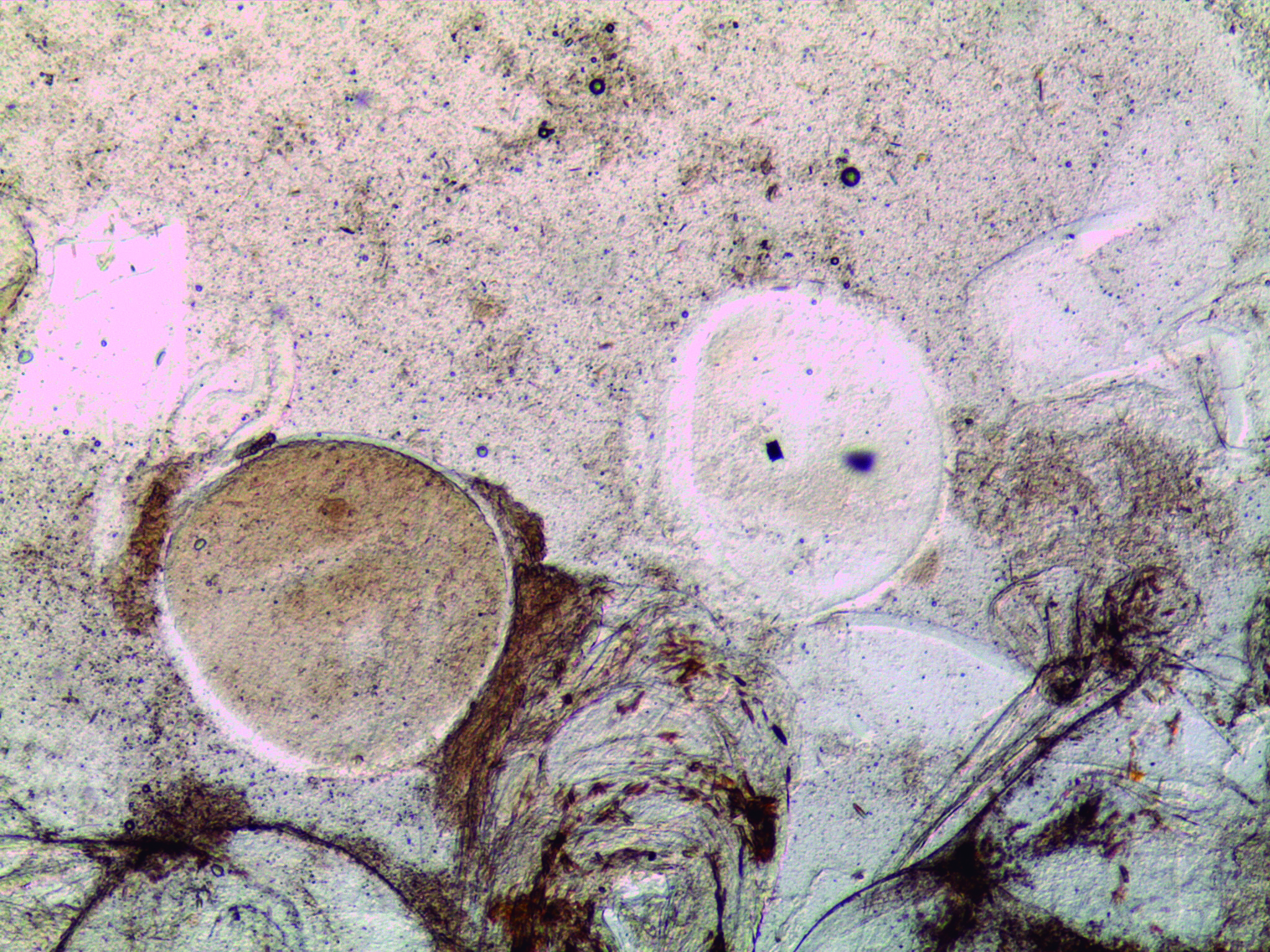
Symptoms: Adult parasites reside in the heart and major blood vessels of the gilthead sea bream. Their eggs travel through the bloodstream to reach the gill capillaries causing capillary rupture and allowing the larvae to exit into the environment and resulting in what is known as “white gill syndrome.” Diagnosis is made by observing eggs in gill capillaries in fresh preparations.
Control: No treatment.
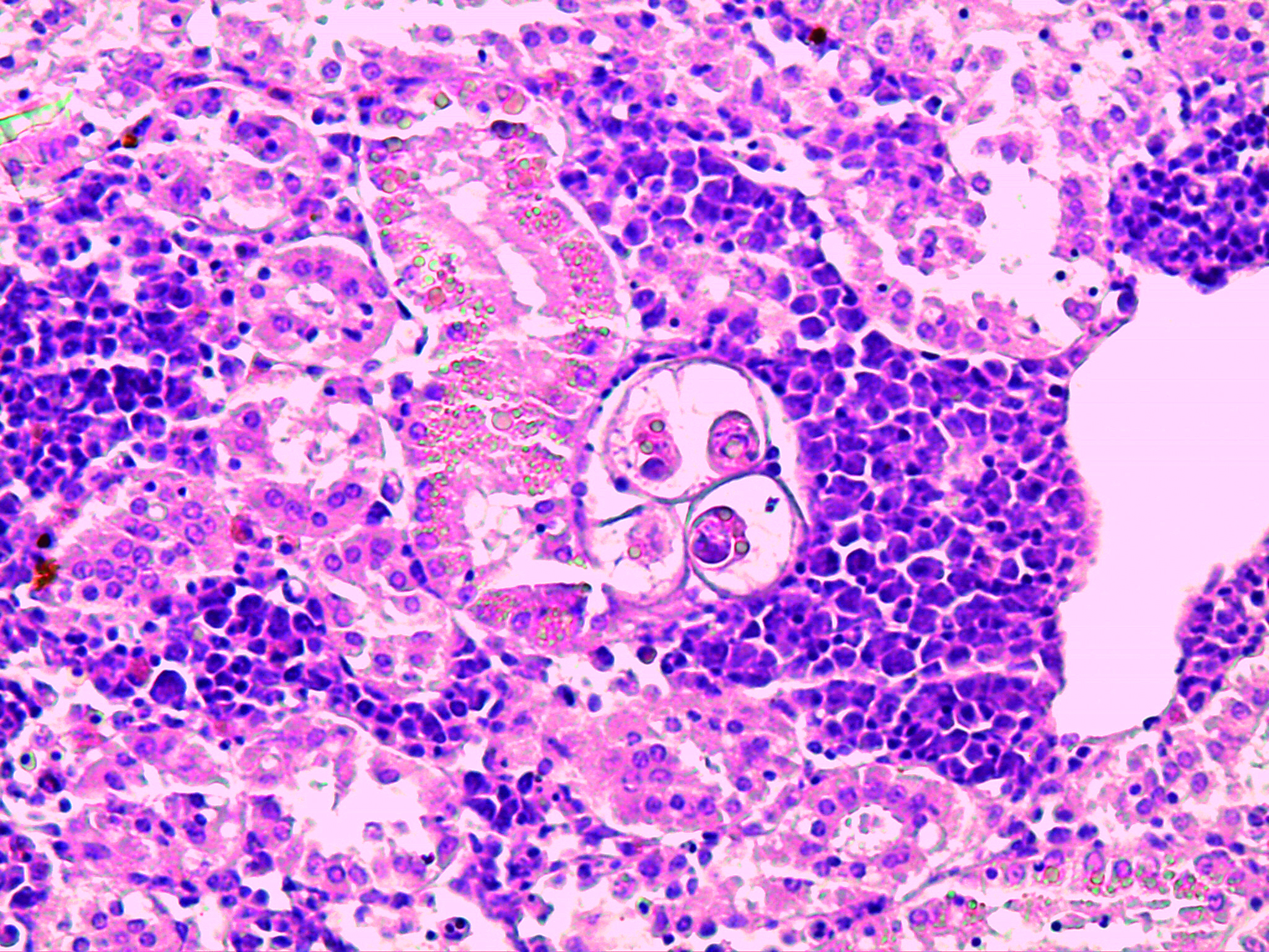
Digestive Microsporidiosis
Agent: Enterospora nucleophila (Enterocytozoonidae, Microsporodian parasite).
Symptoms: Intranuclear protozoan resides within the nuclei of enterocytes and rodlet cells in the digestive tract of gilthead sea bream. Affected fish show emaciation and severe anemia.
It appears during winter, but the disease may become more severe when temperatures rise.
Control: No treatment. Prevention involves separating fish of different sizes to reduce transmission.
Enteromyxosis
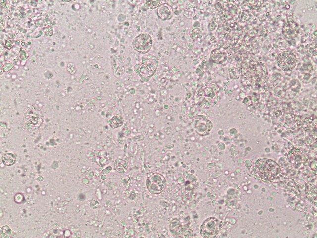
Agent: Enteromyxum leei (Myxidiidae, Myxozoan histozoic parasite).
Symptoms: This Myxosporean parasite infects the intestinal mucosa of gilthead sea bream causing severe weight loss and high mortality. Sea bass may act as a carrier without being affected. Horizontal transmission occurs between fish via pre-sporogonic forms expelled in fecal packets, rather than spores typical of other myxosporeans. Mature spores have a characteristic “croissant” shape with polar capsules at their ends.
Control: No treatment. Prevention involves separating fish of different sizes to reduce transmission, net cleaning and removal of mortalities.
Amyloodiniasis
Agent: Amyloodinium ocellatum (Thoracosphaeraceae, Dinoflagellate parasite).
Symptoms: A dinoflagellate affecting many marine species, primarily in extensive farming systems. Mortality can be high. The life cycle resembles that of Cryptocaryon irritans. Diagnosis involves observing trophonts in fresh gill and skin samples.
Control: Depending on the country regulation, it is possible to use external bath.
Other Endoparasites

- Ceratomyxa sparusaurati, C. diplodae, C. labracis: Coelozoic myxosporeans found in the gallbladder. They generally have little pathological significance.
- Polisporoplasma sparis and Leptotheca sparidarum: Myxosporeans found in the kidney of gilthead sea bream (P. sparis), in parenchyma and in tubules (L. sparidarum). Observed during pre-fattening at high temperatures.
- Cryptosporidium molnari: Found in the digestive tracts of post-larvae and juveniles of sea bream and sea bass, causing growth issues and mortality. Usually resolve over time.
- Pleistophora senegalensis / Glugea sp.: Microsporidians that form xenomas in the muscle tissue of gilthead sea bream.
- Coccidiosis (Eimeria spp.): Intestinal protozoans that can cause severe cachexia in sea bass. Common in estuarine environments.
Other
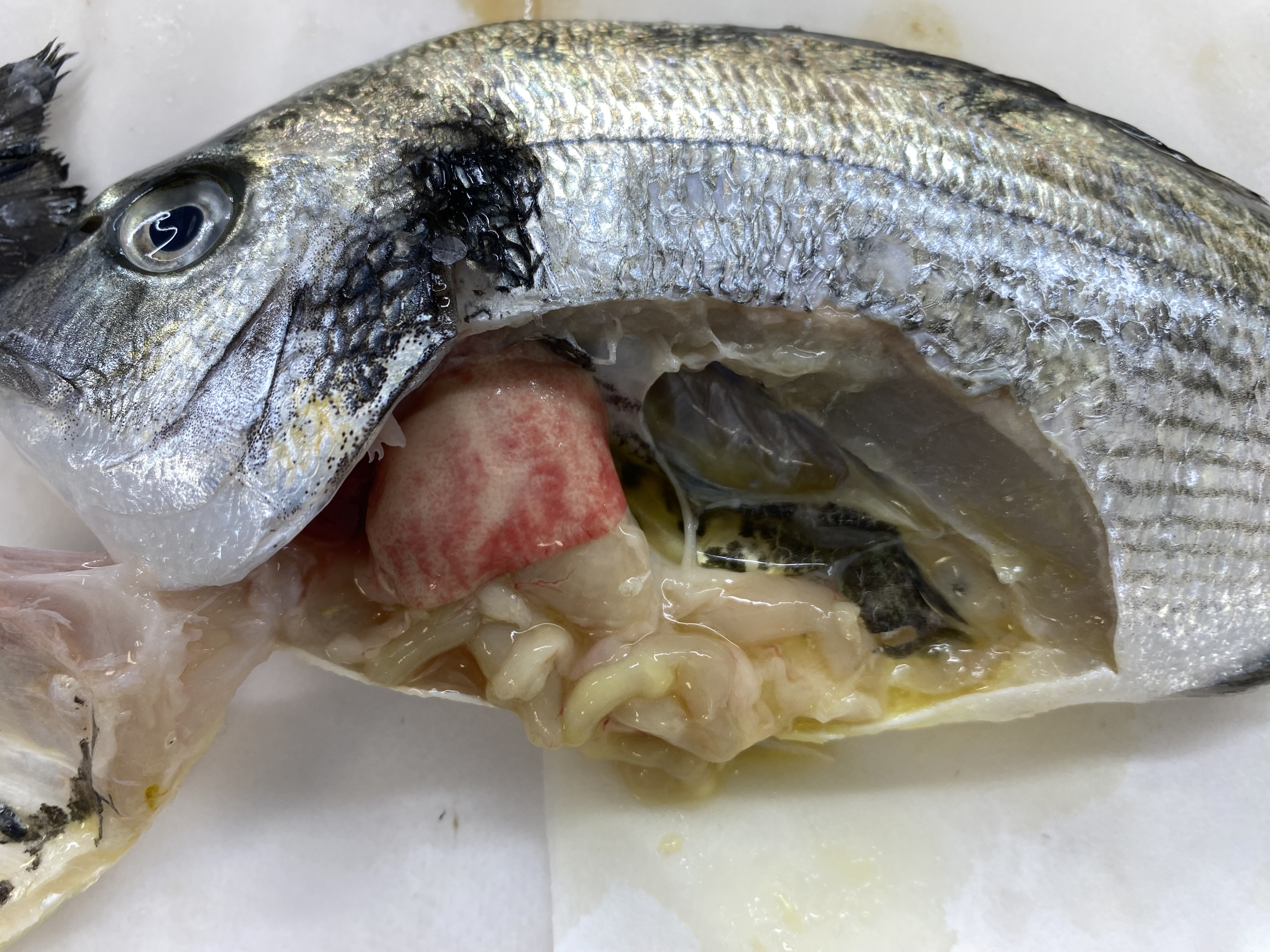
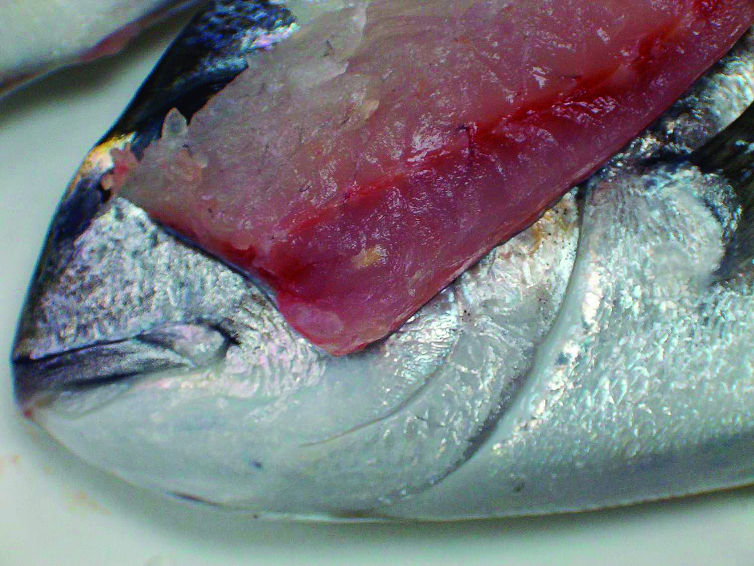
Winter Disease
Agent: Multifactorial issue.
Symptoms: It affects gilthead sea bream from December to April due to cold stress. It courses with erratic swimming, abdominal distension, pale liver, and digestive tract dilation with whitish mucosal cords. Mortality can arise due to combined factors like thermal stress, metabolic imbalances, nutritional deficiencies, and secondary infections. First-year fish experience higher mortality than older fish.
Control: Preventative measures include preparing fish for winter with specialised diets.
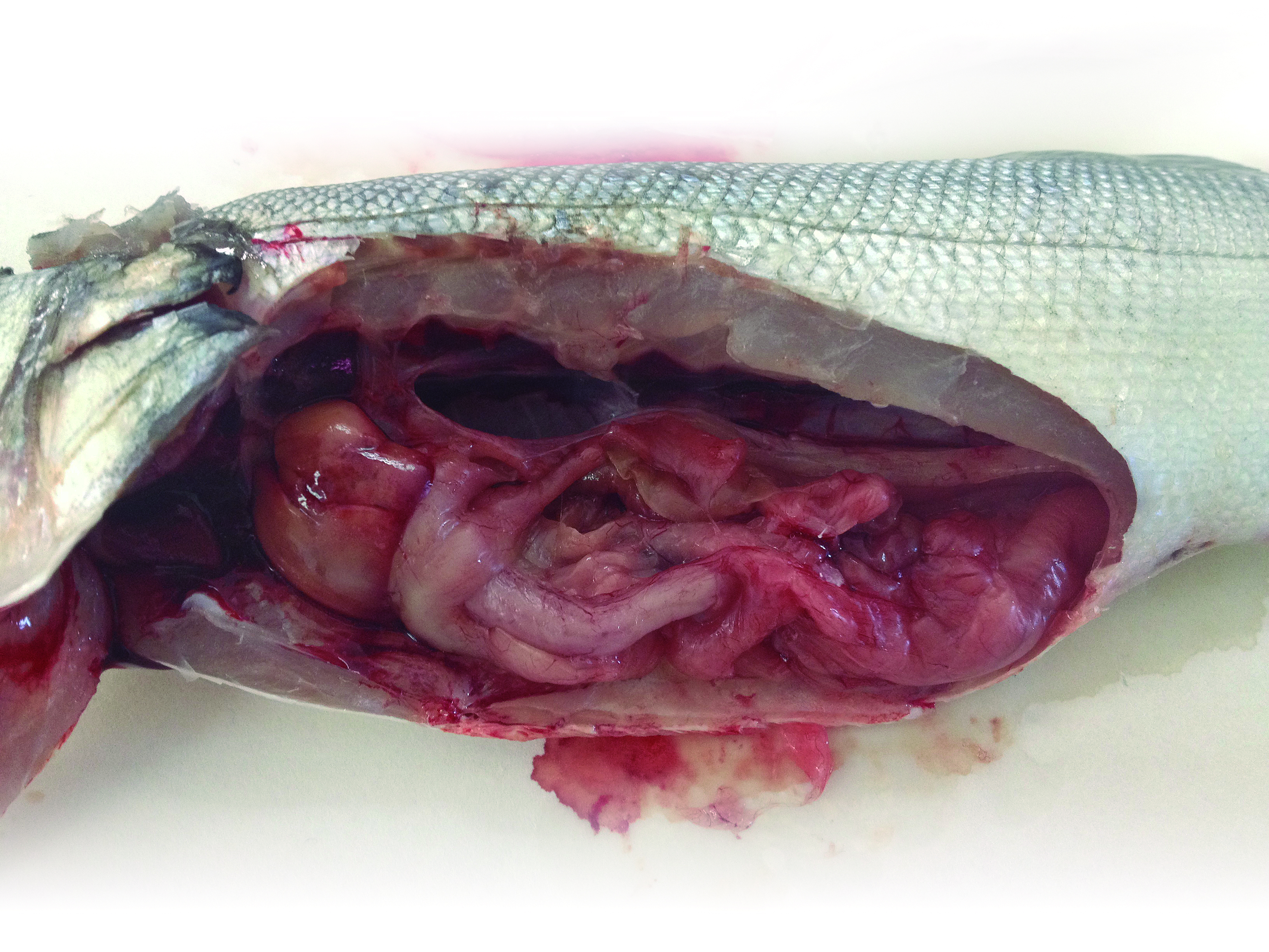
Sudden Death Syndrome
Agent: The cause is unclear but may involve genetic predisposition, farming systems or cardiac issues.
Symptoms: Common in sea bass, associated with sudden stressors (storms, handling, predators). Mortality can occur rapidly, with fish displaying hyperactivity, erratic swimming, and red gills.
Control: Prevention focuses on reducing stress factors.
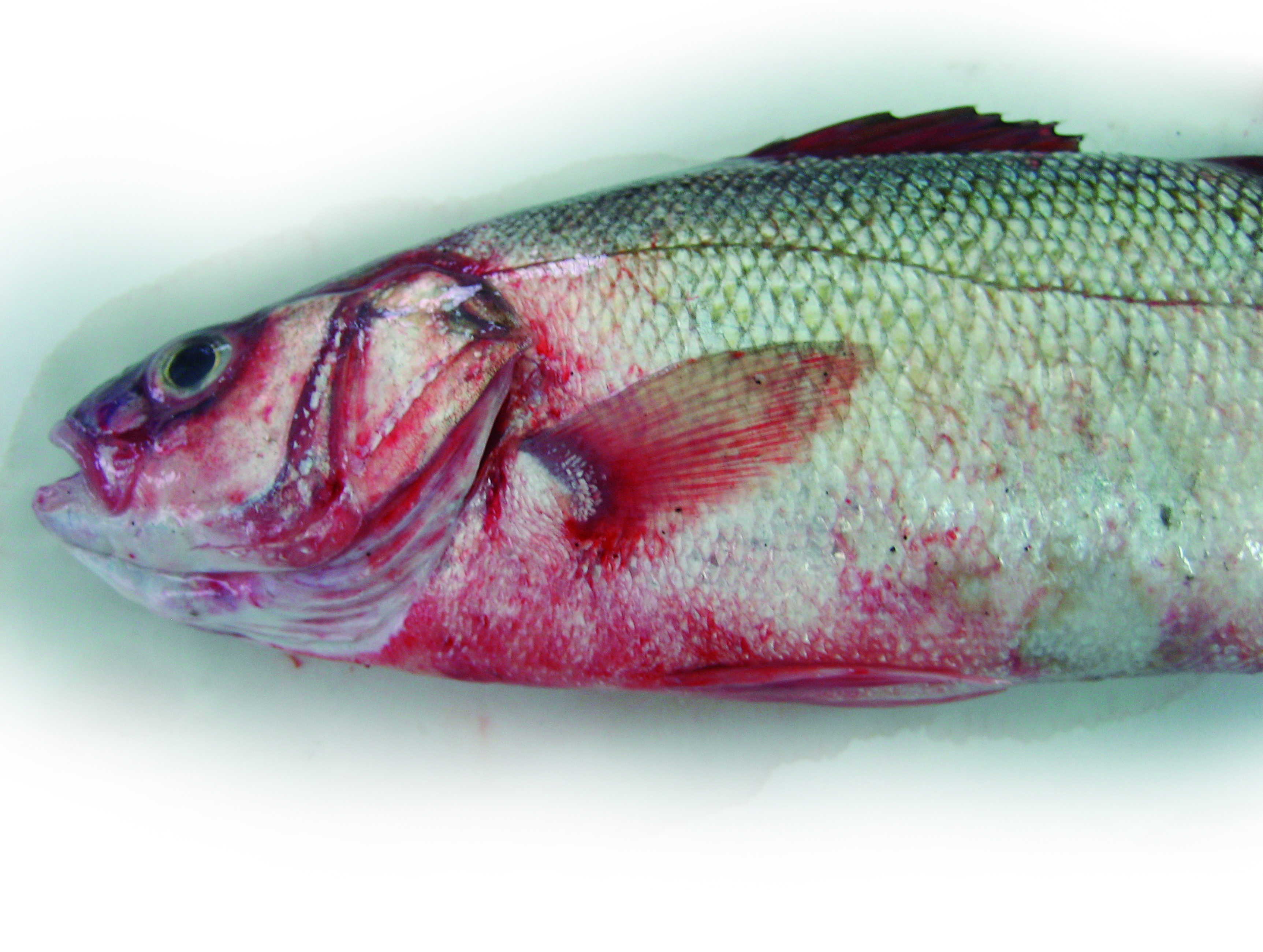
Cutaneous Ulcerative Rash - “Red Spot Syndrome”
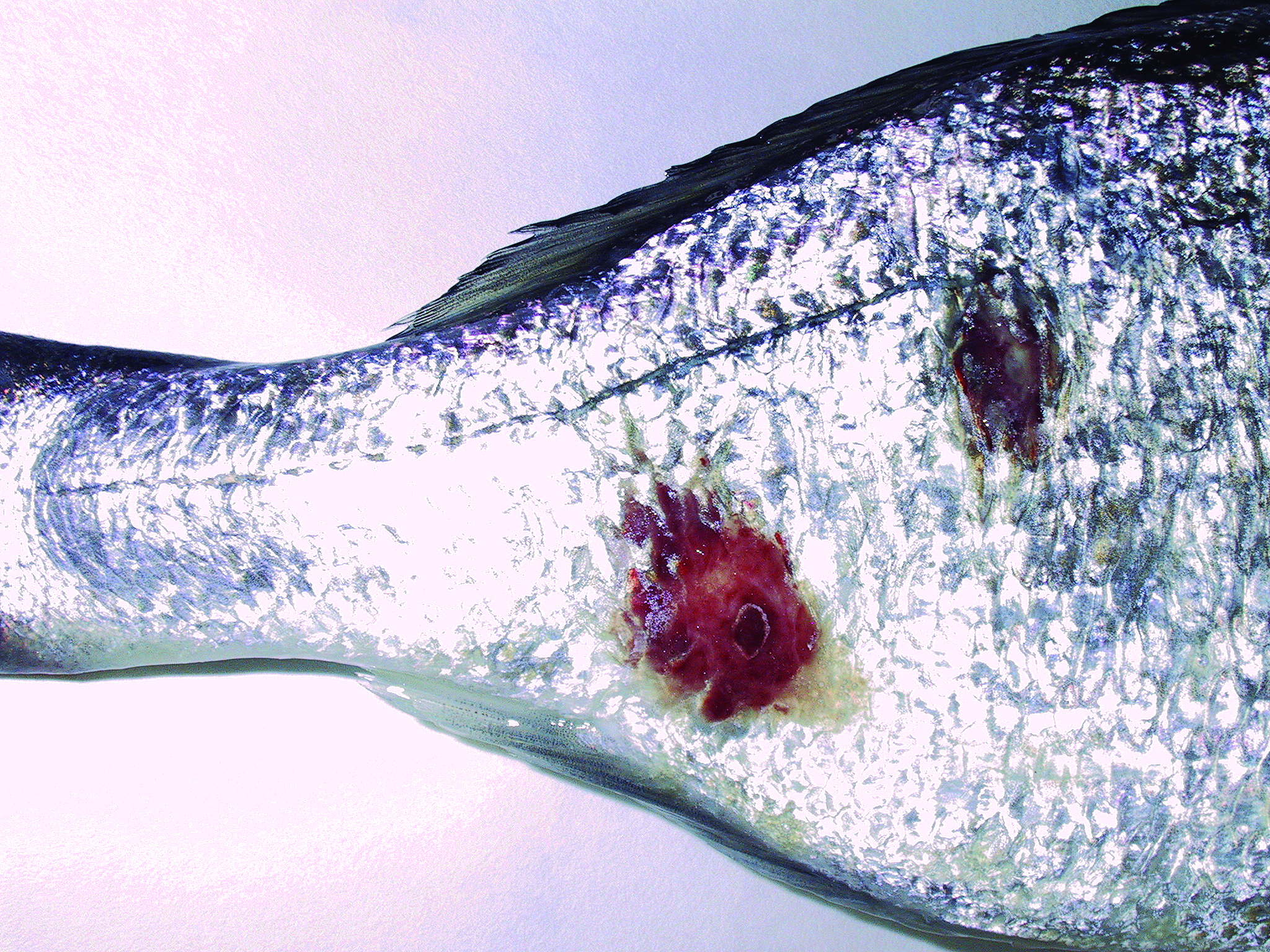
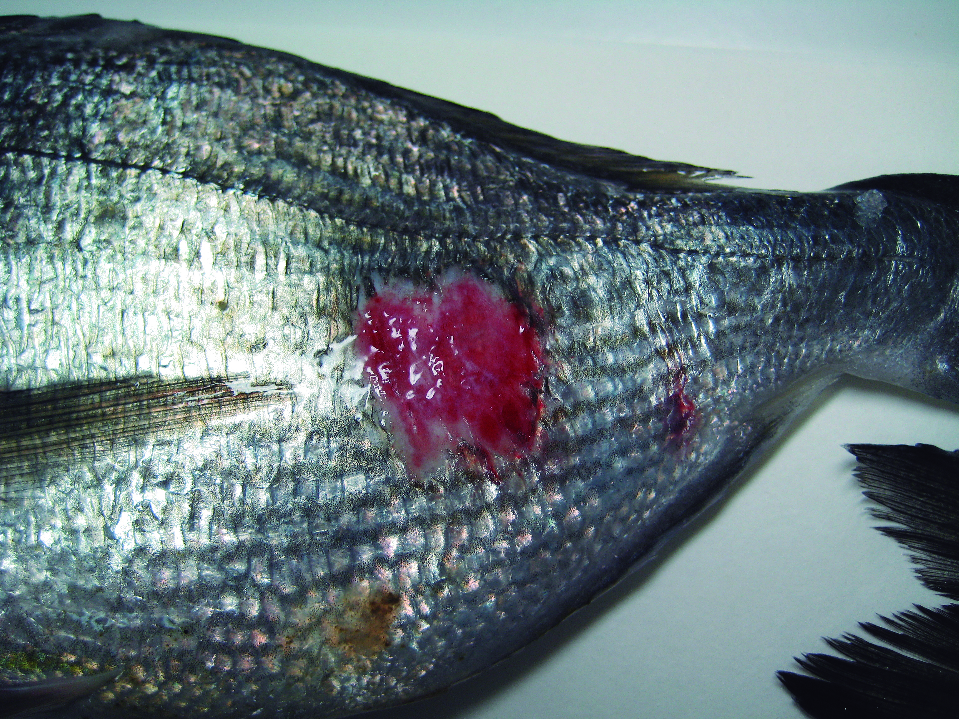
Agent: The cause is unknown but may involve Rickettsia-like organisms (RLOs), allergic reactions, or autoimmune issues.
Symptoms: A disease observed in gilthead sea bream in floating cages, mainly during winter and spring. Fish develop hemorrhagic, erosive and ulcerative lesions that expand. Lesions typically heal as temperatures rise. Morbidity can be high, though mortality is usually low.
Control: Preventative measures include preparing fish for winter with specialized diets.
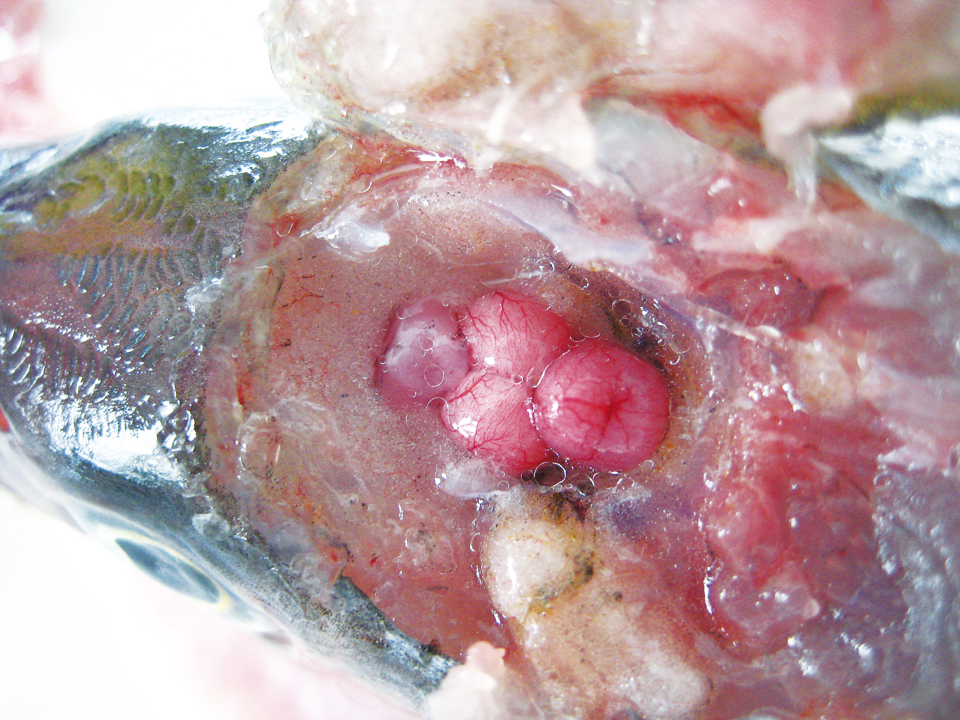
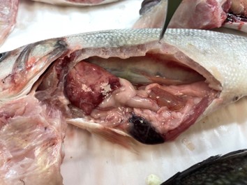
Systemic Granulomas
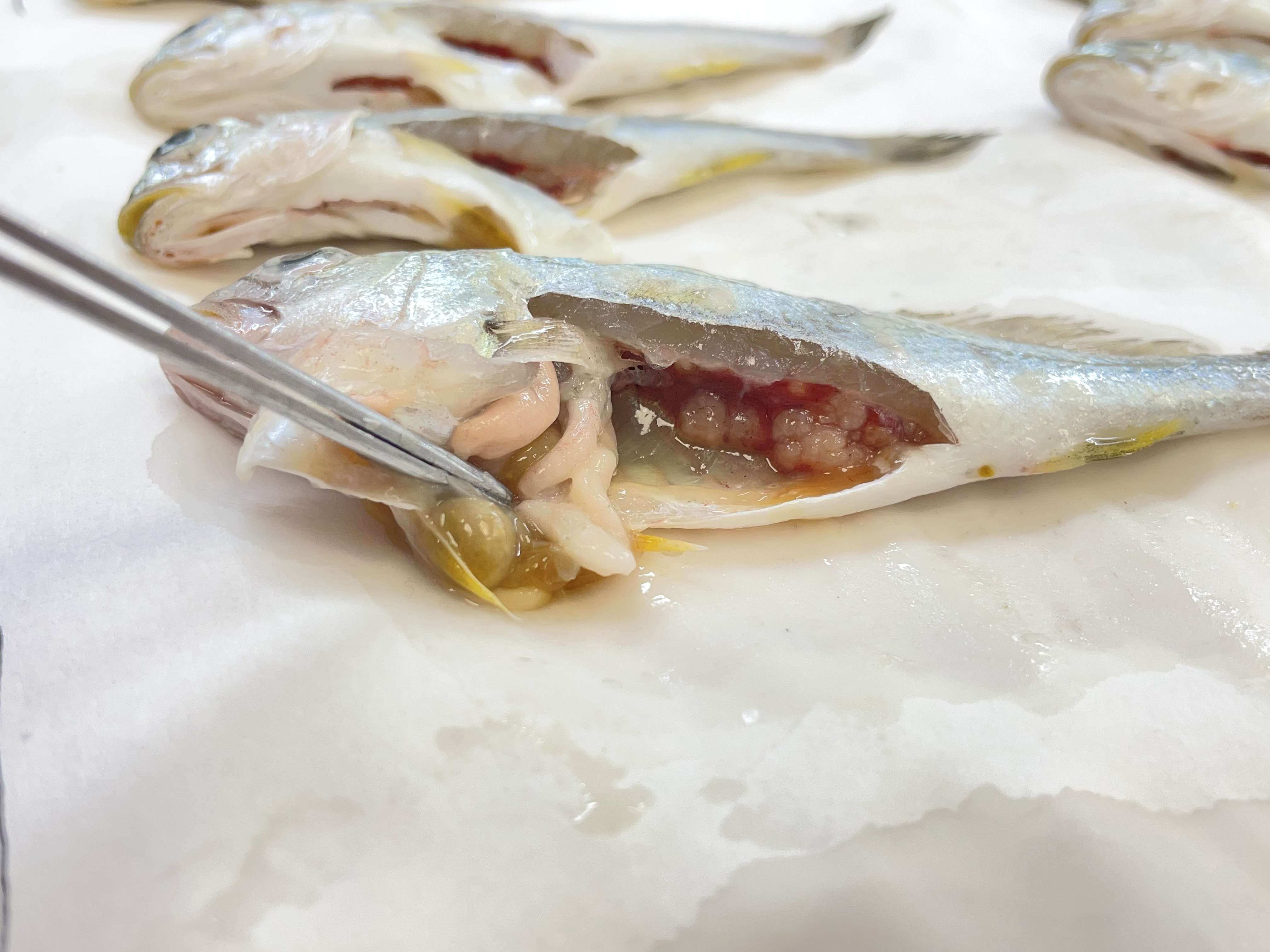
Agent: The cause is unknown. Some potential factors include infectious agents, metabolic issues, nutritional disorders, or autoimmune problems.
Symptoms: Common in meagre (Argyrosomus regius). Fish develop multifocal white nodules visible throughout the viscera, particularly in the kidney, which may become calcified over time. Microscopically, the nodules correspond to granulomas of concentric layers of epithelioid cells around a necrotic core.
Control: No treatment.